KC-0239

293T-CD66e-Cell-Line

×
Please enable JavaScript in your browser to complete this form.
20077
Home » 细胞系 » 293T-CD66e-Cell-Line

Background of 293T-CD66e-Cell-Line

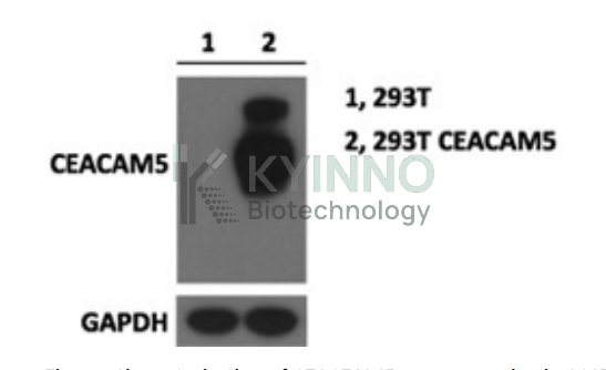
CEACAM-5, also known as CEA and CD66e, belongs to the large family of CEACAM and pregnancy specific glycoproteins. CEACAM-5 is expressed primarily by epithelial cells, consists of an N-terminal Ig-like V-set domain followed by six Ig-like C2-set domains and a GPI anchor. CEACAM-5 functions as a calcium-independent adhesion molecule through homophilic and heterophilic interactions with CEACAM-1. CEACAM-5 is upregulated in a wide variety of human tumors and is a commonly used cancer marker. It promotes tumor cell migration, invasion, adhesion, and metastasis). It also contributes to tumor formation by maintaining cellular proliferation in the presence of differentiation stimuli, and by blocking apoptosis following loss of ECM anchorage (anoikis)

Specifications

Catalog NumberKC-0239
Cell Line Name293T-CD66e-Cell-Line
Host Cell LineHuman HEK293T cell line
DescriptionHEK293T cell line stable expressing exogenous human CEMCAM5 gene
QuantityTwo vials of frozen cells (≥2-106/vial)
StabilityStable in culture over a minimum of 10 passages
ApplicationDrug screening and biological assays
Freezing Medium70% DMEM+20% FBS+10% DMSO
Propagation MediumDMEM+10%FBS+0.5µg/mL Puromycin
Selection MarkerPuromycin
MorphologyEpithelial
SubcultureSplit saturated culture 1:3-1:6 every 3 days; seed out at about 1-3 × 10⁵ cells/mL
Incubation37 °C with 5% CO2
StorageLiquid nitrogen immediately upon receiving
Doubling TimeApproximately 30 hours
Mycoplasma StatusNegative
In Vivo ValidationNA

Cell Line Generation

293T Human CEACAM5 Cell Line was generated using lentiviral Vector with Human CEACAM5 gene sequence.

Characterization

Figure: Characterization of CEACEAM5 overexpressing in 293T stable clones using Western Blot.

Cell Resuscitation

  1. Prewarm culture medium (DMEM + 10% FBS + 0.5μg/mL puromycin)in a 37°C water bath.
  2. Thaw the frozen vial in a 37°C water bath for 1-2 minutes.
  3. Transfer the vial into biosafety cabinet, and wipe the surface with 70% ethanol.
  4. Unscrew the top of the vial and transfer the cell suspension gently into a sterile centrifuge tube containing 9.0mL complete culture medium.
  5. Spin at ~ 125 × g for 5-7 minutes at room temperature, and discard the supernatant without disturbing the pellet.
  6. Resuspend cell pellet with the appropriate volume of complete medium and transfer the cell suspension into a T25 culture flask.
  7. Incubate the flask at 37°C, 5% CO2 incubator.
  8. Split saturated culture 1:3-1:6 every 3 days; seed out at about 1-3 × 10⁵ cells/mL.

Cell Freezing

  1. Prepare the freezing medium (70% DMEM + 20% FBS + 10% DMSO) fresh immediately before use.
  2. Keep the freezing medium on ice and label cryovials.
  3. Transfer cells to a sterile, conical centrifuge tube, and count the cells.
  4. Centrifuge the cells at 250×g for 5 minutes at room temperature and carefully aspirate off the medium.
  5. Resuspend the cells at a density of at least 3×10⁶ cells/mL in chilled freezing medium.
  6. Aliquot 1 mL of the cell suspension into each cryovial.
  7. Freeze cells in the CoolCell freezing container overnight in a -80°C freezer.
  8. Transfer vials to liquid nitrogen for long-term storage

References

  1. Oikawa S, Nakazato H, Kosaki G (1987). "Primary structure of human carcinoembryonic antigen (CEA) deduced from cDNA sequence". Biochem. Biophys. Res. Commun. 142(2): 511ÿ8.
  2. Wirth T, Soeth E, Czubayko F, Juhl H (2002). "Inhibition of endogenous carcinoembryonic antigen (CEA) increases the apoptotic rate of colon cancer cells and inhibits metastatic tumor growth". Clin. Exp. Metastasis. 19 (2): 155ÿ60.
Please enable JavaScript in your browser to complete this form.