KC-1047

A549-OS8 Cell Line

×
Please enable JavaScript in your browser to complete this form.
21439
Home » 细胞系 » A549-OS8 Cell Line

Background of A549-OS8 Cell Line

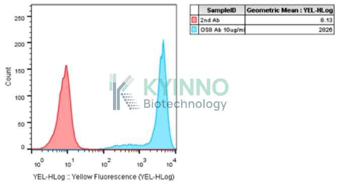
The OS8 antigen is a cell surface protein identified as a potential tumor-associated antigen. It is highly expressed in certain cancers, most notably osteosarcoma (as suggested by its name), making it a candidate for targeted immunotherapy research. Its expression profile in malignancies compared to limited expression in normal tissues positions it as a promising target for the development of antibody-based therapies, such as monoclonal antibodies or CAR-T cells, aimed at treating solid tumors.

Specifications

Catalog NumberKC-1047
Cell Line NameA549-OS8 Cell Line
Clone NumberNA
Host Cell LineA549
DescriptionStable A549 cell line expressing exogenous OS8 gene
QuantityTwo vials of frozen cells (≥2-106/vial)
StabilityStable in culture over a minimum of 10 passages
ApplicationDrug screening and biological assays
Freezing Medium70% McCoy’s 5a + 20% FBS + 10% DMSO
Propagation MediumMcCoy's 5a+10%FBS+500µg/mL Hygromycin B
Selection MarkerHygromycin B
MorphologyEpithelial
SubcultureSplit saturated culture 1:4-1:8 every 2-3 days; seed out at about 1-3 × 10⁵ cells/mL
Incubation37 °C with 5% CO2
StorageLiquid nitrogen immediately upon receiving
Doubling TimeApproximately 22 hours
Mycoplasma StatusNegative
In Vivo ValidationNA

Cell Line Generation

A549-OS8 Cell Line was generated using a lentiviral vector expressing the OS8 sequence.

Characterization

Figure 1: Characterization of OS8 overexpression in the A549-OS8 stable clone using FACS.

Cell Resuscitation

  1. Prewarm culture medium (McCoy's 5a+10%FBS+500µg/mL Hygromycin B)in a 37°C water bath.
  2. Thaw the frozen vial in a 37°C water bath for 1-2 minutes.
  3. Transfer the vial into biosafety cabinet, and wipe the surface with 70% ethanol.
  4. Unscrew the top of the vial and transfer the cell suspension gently into a sterile centrifuge tube containing 9.0mL complete culture medium.
  5. Spin at ~ 125 × g for 5-7 minutes at room temperature, and discard the supernatant without disturbing the pellet.
  6. Resuspend cell pellet with the appropriate volume of complete medium and transfer the cell suspension into a T25 culture flask.
  7. Incubate the flask at 37°C, 5% CO2 incubator.
  8. Split saturated culture 1:4-1:8 every 2-3 days; seed out at about 1-3 × 10⁵ cells/mL.

Cell Freezing

  1. Prepare the freezing medium (70% McCoy’s 5a + 20% FBS + 10% DMSO) fresh immediately before use.
  2. Keep the freezing medium on ice and label cryovials.
  3. Transfer cells to a sterile, conical centrifuge tube, and count the cells.
  4. Centrifuge the cells at 250×g for 5 minutes at room temperature and carefully aspirate off the medium.
  5. Resuspend the cells at a density of at least 3×10⁶ cells/mL in chilled freezing medium.
  6. Aliquot 1 mL of the cell suspension into each cryovial.
  7. Freeze cells in the CoolCell freezing container overnight in a -80°C freezer.
  8. Transfer vials to liquid nitrogen for long-term storage.

References

  1. Latouche, Jean-Baptiste; Sadelain, Michel (2000). "Induction of human cytotoxic T lymphocytes by artificial antigen-presenting cells". Nature Biotechnology. 18 (4): 405–409.
  2. Perica, Karlo; Kosmides, Alyssa K; Schneck, Jonathan P (2015). "Linking form to function: Biophysical aspects of artificial antigen presenting cell design". Biochimica et Biophysica Acta (BBA) - Molecular Cell Research. 1853 (4): 781–790.
Please enable JavaScript in your browser to complete this form.