KC-1069

CHOK1-FCGR2A-R167-Cell-Line

×
Please enable JavaScript in your browser to complete this form.
21477
Home » CHOK1-FCGR2A-R167-Cell-Line

Background of CHOK1-FCGR2A-R167-Cell-Line

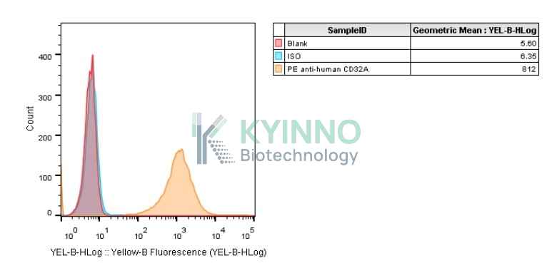
FCGR2A, also known as CD32A, is the low-affinity receptor for the immunoglobulin G Fc fragment that is highly expressed on myeloid cells and expressed on a small subset of T cells. The protein encoded by this gene is a cell surface receptor found on phagocytic cells such as macrophages and neutrophils, and is involved in the process of phagocytosis and clearing of immune complexes. FCGR2A was proposed by Descours et al. as a marker of HIV reservoir cells. Diseases associated with FCGR2A include Cystic Fibrosis and Systemic Lupus Erythematosus.

Specifications

Catalog NumberKC-1069
Cell Line NameCHOK1-FCGR2A-R167-Cell-Line
Clone Number1#
Host Cell LineChinese hamster ovary CHO-k1 cell line
DescriptionStable CHO-K1 cell line expressing exogenous FCGR2A R167 gene
QuantityTwo vials of frozen cells (≥2-106/vial)
StabilityStable in culture over a minimum of 10 passages
ApplicationDrug screening and biological assays
Freezing Medium70% RPMI1640+20% FBS+10% DMSO
Propagation MediumRPMI1640 + 10% FBS + 10μg/ml Puromycin
Selection MarkerPuromycin
MorphologyEpithelial
SubcultureSplit saturated culture 1:4-1:8 every 2-3 days; seed out at about 1-2 × 10⁵ cells/mL
Incubation37 °C with 5% CO2
StorageLiquid nitrogen immediately upon receiving
Doubling TimeApproximately 24 hours
Mycoplasma StatusNegative
In Vivo ValidationNA

Cell Line Generation

CHO-K1 FCGR2A R167 Cell Line was generated using a lentiviral vector expressing the FCGR2A R167 sequence.

Characterization

Figure1: Characterization of FCGR2A overexpression in CHO-K1 stable clones using FACS.

Figure2: Characterization of CHO-K1 FCGR2A R167 Cell Line stable clone using PCR sequencing.

Cell Resuscitation

1. Prewarm culture medium (RPMI1640 supplemented with 10% FBS and 10μg/mL Puromycin)in a 37°C water bath.
2. Thaw the frozen vial in a 37°C water bath for 1-2 minutes.
3. Transfer the vial into biosafety cabinet, and wipe the surface with 70% ethanol.
4. Unscrew the top of the vial and transfer the cell suspension gently into a sterile centrifuge tube containing 9.0mL complete culture medium.
5. Spin at ~ 125 × g for 5-7 minutes at room temperature, and discard the supernatant without disturbing the pellet.
6. Resuspend cell pellet with the appropriate volume of complete medium and transfer the cell suspension into a T25 culture flask.
7. Incubate the flask at 37°C, 5% CO2 incubator.
8. Split saturated culture 1:4-1:8 every 2-3 days; seed out at about 1-2 × 10⁵ cells/mL.

Cell Freezing

1. Prepare the freezing medium (70% RPMI1640 + 20% FBS + 10% DMSO) fresh immediately before use.
2. Keep the freezing medium on ice and label cryovials.
3. Transfer cells to a sterile, conical centrifuge tube, and count the cells.
4. Centrifuge the cells at 250×g for 5 minutes at room temperature and carefully aspirate off the medium.
5. Resuspend the cells at a density of at least 3×10⁶ cells/mL in chilled freezing medium.
6. Aliquot 1 mL of the cell suspension into each cryovial.
7. Freeze cells in the CoolCell freezing container overnight in a -80°C freezer.
8. Transfer vials to liquid nitrogen for long-term storage

References

  1. Anania JC, Chenoweth AM, Wines BD, Hogarth PM (2019). "The Human FcÎÛRII (CD32) Family of Leukocyte FcR in Health and Disease". Frontiers in Immunology. 10: 464. doi:10.3389/fimmu.2019.00464. PMC 6433993. PMID 30941127.
  2. Hamzeh-Cognasse H, Damien P, Chabert A, Pozzetto B, Cognasse F, Garraud O (2015-02-26). "Platelets and infections - complex interactions with bacteria". Frontiers in Immunology. 6: 82. doi:10.3389/fimmu.2015.00082. PMC 4341565. PMID 25767472.
Please enable JavaScript in your browser to complete this form.