KC-1510

Jurkat-NFAT-Luc2-LAG3-CD3zeta

×
请在浏览器中启用JavaScript来完成此表单。
22281
Home » Jurkat-NFAT-Luc2-LAG3-CD3zeta

Background of Jurkat-NFAT-Luc2-LAG3-CD3zeta

"Lymphocyte-activation gene 3(LAG3), also known as CD223, is a member of immunoglobulin superfamily, expressed on activated T cells, NK cells, B cells and DC cells. LAG3 can bind to MHC II molecules and induce maturation of DC cells, and cytokine secretion of cytotoxic T cells and NK cells. LAG3 is a promising drug target of cancer therapy and autoimmune disease, its therapeutic antibodies including BMS-986016 and GSK2831781 had already been used in the clinical trial. NFAT (nuclear factor of activator T cells) proteins belongs to a family of transcription factors that are very important in T cells activation, differentiation and tolerance. NFAT pathway is activated by Ca2+ and the Ca2+/calmodulin- dependent serine phosphatase calcineurin after the engagement of T cell receptor (TCR)."

Specifications

Catalog NumberKC-1510
Cell Line NameJurkat-NFAT-Luc2-LAG3-CD3zeta
Host Cell LineJurkat-NFAT-Luc2
DescriptionJurkat cell line stable expressing exogenous luciferase gene under the control of NFAT responsive element and full-length human LAG3-CD3zeta fusion gene
QuantityOne vial of frozen cells (≥2-106/vial)
StabilityStable in culture over a minimum of 10 passages
ApplicationDrug screening and biological assays
Freezing Medium70% RPMI1640 + 20% FBS + 10% DMSO
Propagation MediumRPMI1640 + 10% FBS + 300μg/mL Hygromycin + 0.75μg/mL puromycin
Selection MarkerHygromycin B and puromycin
MorphologyLymphoblast
SubcultureSplit saturated culture 1:4-1:5 every 2-3 days; seed out at about 1 × 10⁵ cells/mL
Incubation37 °C with 5% CO2
StorageLiquid nitrogen immediately upon receiving
Doubling TimeApproximately 30 hours
Mycoplasma StatusNegative
In Vivo ValidationNA

Cell Line Generation

Jurkat-NFAT-Luc2-LAG3-CD3zeta cell line was generated using lentivirus vector expressing LAG3-CD3zeta fusion sequence.

Characterization

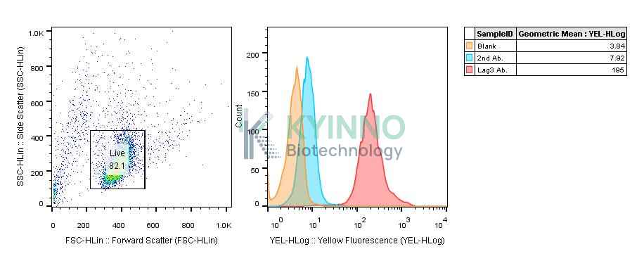
Figure 1: Characterization of human LAG3 overexpression in Jurkat-NFAT-Luc2-LAG3-CD3zeta stable clones using FCAS.

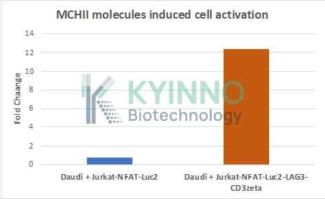
Figure 2: Jurkat-NFAT-Luc2-LAG3-CD3zeta cells were seeded into the 96-well plate, treated with Daudi for 16 hours, and then readout using Bright-Glo Detection System.

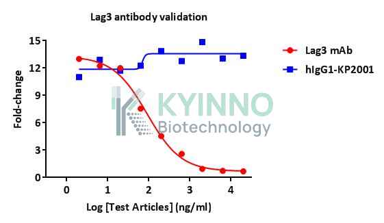
Figure 3: Jurkat-NFAT-Luc2-LAG3-CD3zeta cells were seeded into the 96-well plate, treated with Lag3 mAb for 16 hours, and then readout using Bright-Glo Detection System.

Cell Resuscitation

  1. Prewarm culture medium (RPMI1640 supplemented with 10% FBS, 300μg/mL Hygromycin B and 0.75μg/mL puromycin).
  2. Thaw the frozen vial in a 37°C water bath for 1-2 minutes.
  3. Transfer the vial into biosafety cabinet, and wipe the surface with 70% ethanol.
  4. Unscrew the top of the vial and transfer the cell suspension gently into a sterile centrifuge tube containing 9.0mL complete culture medium.
  5. Spin at ~ 125 × g for 5-7 minutes at room temperature, and discard the supernatant without disturbing the pellet.
  6. Resuspend cell pellet with the appropriate volume of complete medium and transfer the cell suspension into a T25 culture flask.
  7. Incubate the flask at 37°C, 5% CO2 incubator.
  8. Split saturated culture 1:4-1:8 every 2-3 days; seed out at about 1-3 × 10⁵ cells/mL.

Cell Freezing

  1. Prepare the freezing medium (70% RPMI1640 + 20% FBS + 10% DMSO) fresh immediately before use.
  2. Keep the freezing medium on ice and label cryovials.
  3. Transfer cells to a sterile, conical centrifuge tube, and count the cells.
  4. Centrifuge the cells at 250×g for 5 minutes at room temperature and carefully aspirate off the medium.
  5. Resuspend the cells at a density of at least 3×10⁶ cells/mL in chilled freezing medium.
  6. Aliquot 1 mL of the cell suspension into each cryovial.
  7. Freeze cells in the CoolCell freezing container overnight in a -80°C freezer.
  8. Transfer vials to liquid nitrogen for long-term storage

References

  1. Anderson, Ana C, Nicole Joller, and Vijay K Kuchroo. 2016. “Lag-3, Tim-3, and TIGIT: Co-Inhibitory Receptors with Specialized Functions in Immune Regulation.” Immunity 44 (5). Elsevier Inc.: 989–1004.
  2. Huard, B, R Mastrangeli, P Prigent, D Bruniquel, S Donini, N El-Tayar, B Maigret, M Dréano, and F Triebel. 1997. “Characterization of the Major Histocompatibility Complex Class II Binding Site on LAG-3 Protein.” Proceedings of the National Academy of Sciences 94 (11): 5744–49.

Use License Agreement

Research Use Only.
Not for use in diagnostic procedures or therapeutic applications.
Redistribution of the cell line or its derivatives is prohibited without prior written permission from Kyinno Biotechnology.
请在浏览器中启用JavaScript来完成此表单。