KC-1667

293T-NFκB-Luc2-mouse-CD40-reporter-Cell-Line

×
Please enable JavaScript in your browser to complete this form.
22577
Home » 293T-NFκB-Luc2-mouse-CD40-reporter-Cell-Line

Background of 293T-NFκB-Luc2-mouse-CD40-reporter-Cell-Line

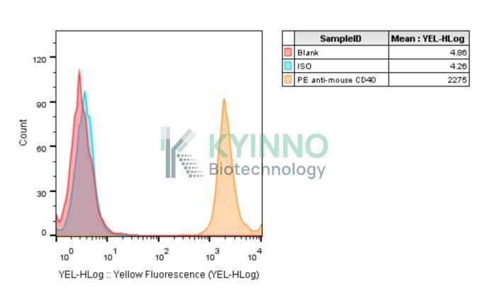
CD40, the receptor for CD40L, is a member of the TNF receptor (TNFR) superfamily. It is expressed in a plethora of cell types, including B cells, macrophages and dendritic, endothelial and epithelial cells.The CD40/CD40L interactions play a central role in orchestrating the immune response. CD40L is produced by both Th1 and Th2 helper T cells, as well as by mast cells, basophils and eosinophils.Signals induced by CD40L expressed in Th1 and Th2 cells are required for macrophage and B‐cell activation, respectively.It transduces TRAF6- and MAP3K8-mediated signals that activate ERK in macrophages and B cells, leading to induction of immunoglobulin secretion.

Specifications

Catalog NumberKC-1667
Cell Line Name293T-NFκB-Luc2-mouse-CD40-reporter-Cell-Line
Host Cell Line293T-NFκB-Luc2
DescriptionStable 293T cell line expressing exogenous luciferase gene under the control of CD40 signaling pathway.
QuantityTwo vials of frozen cells (≥2-106/vial)
StabilityStable in culture over a minimum of 10 passages
ApplicationCell model for monitoring mouse CD40 signaling pathway
Freezing Medium70% DMEM+20% FBS+10% DMSO
Propagation MediumDMEM+10% FBS +150μg/ml Hygromycin B+1μg/ml puromycin
Selection MarkerHygromycin B and Puromycin
MorphologyEpithelial
SubcultureSplit saturated culture 1:4-1:8 every 2-3 days; seed out at about 1-3 × 10⁵ cells/mL
Incubation37 °C with 5% CO2
StorageLiquid nitrogen immediately upon receiving
Doubling TimeApproximately 30 hours
Mycoplasma StatusNegative
In Vivo ValidationNA

Cell Line Generation

293T-NFκB-Luc2-mouse-CD40-reporter cell line was generated using lentivirus expressing mouse CD40 sequence.

Characterization

Figure 1. Characterization of mouse-CD40 overexpression in the 293T-NFκB-Luc2-mouse-CD40 stable clone using FACS.

Cell Resuscitation

  1. Prewarm culture medium (DMEM supplemented with 10% FBS, 150μg/ml Hygromycin and 1μg/ml Puromycin)in a 37°C water bath.
  2. Thaw the frozen vial in a 37°C water bath for 1-2 minutes.
  3. Transfer the vial into biosafety cabinet, and wipe the surface with 70% ethanol.
  4. Unscrew the top of the vial and transfer the cell suspension gently into a sterile centrifuge tube containing 9.0mL complete culture medium.
  5. Spin at ~ 125 × g for 5-7 minutes at room temperature, and discard the supernatant without disturbing the pellet.
  6. Resuspend cell pellet with the appropriate volume of complete medium and transfer the cell suspension into a T25 culture flask.
  7. Incubate the flask at 37°C, 5% CO2 incubator.
  8. Split saturated culture 1:4-1:8 every 2-3 days; seed out at about 1-3 × 10⁵ cells/mL.

Cell Freezing

  1. Prepare the freezing medium (70% DMEM + 20% FBS + 10% DMSO) fresh immediately before use.
  2. Keep the freezing medium on ice and label cryovials.
  3. Transfer cells to a sterile, conical centrifuge tube, and count the cells.
  4. Centrifuge the cells at 250×g for 5 minutes at room temperature and carefully aspirate off the medium.
  5. Resuspend the cells at a density of at least 3×10⁶ cells/mL in chilled freezing medium.
  6. Aliquot 1 mL of the cell suspension into each cryovial.
  7. Freeze cells in the CoolCell freezing container overnight in a -80°C freezer.
  8. Transfer vials to liquid nitrogen for long-term storage

References

  1. Eliopoulos AG, Wang CC, Dumitru CD, Tsichlis PN. Tpl2 transduces CD40 and TNF signals that activate ERK and regulates IgE induction by CD40. EMBO J. 2003 Aug 1;22(15):3855-64. doi: 10.1093/emboj/cdg386. PMID: 12881420; PMCID: PMC169059.
  2. Karnell JL, Rieder SA, Ettinger R, Kolbeck R. Targeting the CD40-CD40L pathway in autoimmune diseases: Humoral immunity and beyond. Adv Drug Deliv Rev. 2019 Feb 15;141:92-103. doi: 10.1016/j.addr.2018.12.005. Epub 2018 Dec 13. PMID: 30552917.
Please enable JavaScript in your browser to complete this form.