KC-1902

293T-SMAD-Luc2 Cell Line

×
请在浏览器中启用JavaScript来完成此表单。
23019
Home » 293T-SMAD-Luc2 Cell Line

Background of 293T-SMAD-Luc2 Cell Line

Transforming growth factor-β1 (TGF-β1) is considered as a crucial mediator in tissue fibrosis and causes tissue scarring largely by activating its downstream small mother against decapentaplegic (Smad) signaling. Different TGF-β signalings play different roles in fibrogenesis. TGF-β1 directly activates Smad signaling which triggers pro-fibrotic gene overexpression. Smad transcription factors lie at the core of one of the most versatile cytokine signaling pathways in metazoan biology-the transforming growth factor-beta (TGFbeta) pathway. Recent progress has shed light into the processes of Smad activation and deactivation, nucleocytoplasmic dynamics, and assembly of transcriptional complexes. A rich repertoire of regulatory devices exerts control over each step of the Smad pathway.

Specifications

Catalog NumberKC-1902
Cell Line Name293T-SMAD-Luc2 Cell Line
Host Cell Line293T
DescriptionStable 293T cell line expressing exogenous luciferase gene under the control of SMAD transcription factors
QuantityOne vial of frozen cells (≥2-106/vial)
StabilityStable in culture over a minimum of 10 passages
ApplicationDrug screening and biological assays
Freezing Medium70% DMEM+20% FBS+10% DMSO
Propagation MediumDMEM+10% FBS +150μg/ml Hygromycin B
Selection MarkerHygromycin B
MorphologyEpithelial
SubcultureSplit saturated culture 1:4-1:8 every 2-3 days; seed out at about 1-3 × 10⁵ cells/mL
Incubation37 °C with 5% CO2
StorageLiquid nitrogen immediately upon receiving
Doubling TimeApproximately 30 hours
Mycoplasma StatusNegative
In Vivo ValidationNA

Cell Line Generation

293T-SMAD-Luc2 cell line was generated using lentivirus expressing SMAD sequence.

Characterization

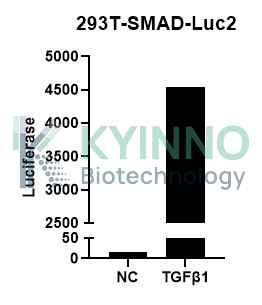
Figure 1. 293T-SMAD-Luc2 cell line was seeded into the 96-well plate, and treated with TGFβ1 at a maximum concentration of 10ng/mL for 6 hours, then readout with Bright-Glo system.

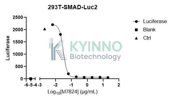
Figure 2. 293T-SMAD-Luc2 cell line was seeded into the 96-well plate, and treated with TGFβ1 and M7824 at a maximum concentration of 10ng/mL and 20μg/mL for 6 hours, then readout with Bright-Glo system.

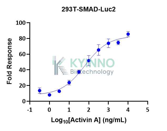
Figure 3. 293T-SMAD-Luc2 cell line was seeded into the 96-well plate, and treated with Activin A at a maximum concentration of 10μg/mL for 6 hours, then readout with Bright-Glo system.

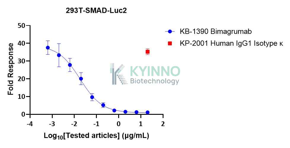
Figure 4. 293T-SMAD-Luc2 cell line was seeded into the 96-well plate, and treated with Activin A and Bimagrumab at a maximum concentration of 50ng/mL and 20μg/mL for 6 hours, then readout with Bright-Glo system.

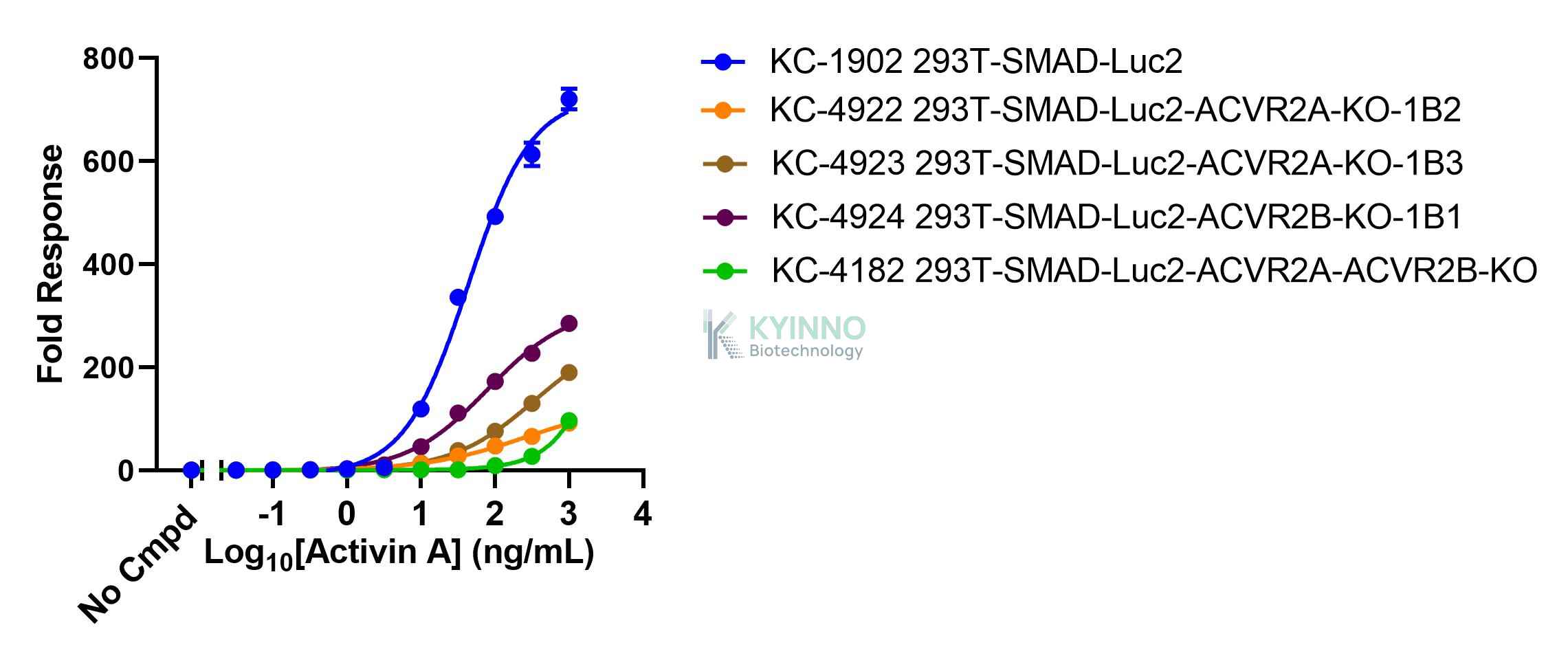
Figure 5. 293T-SMAD-Luc2 cells were seeded into 96-well plates, treated with Activin A for 16 hours, and then read out using Bright-Glo Detection System.

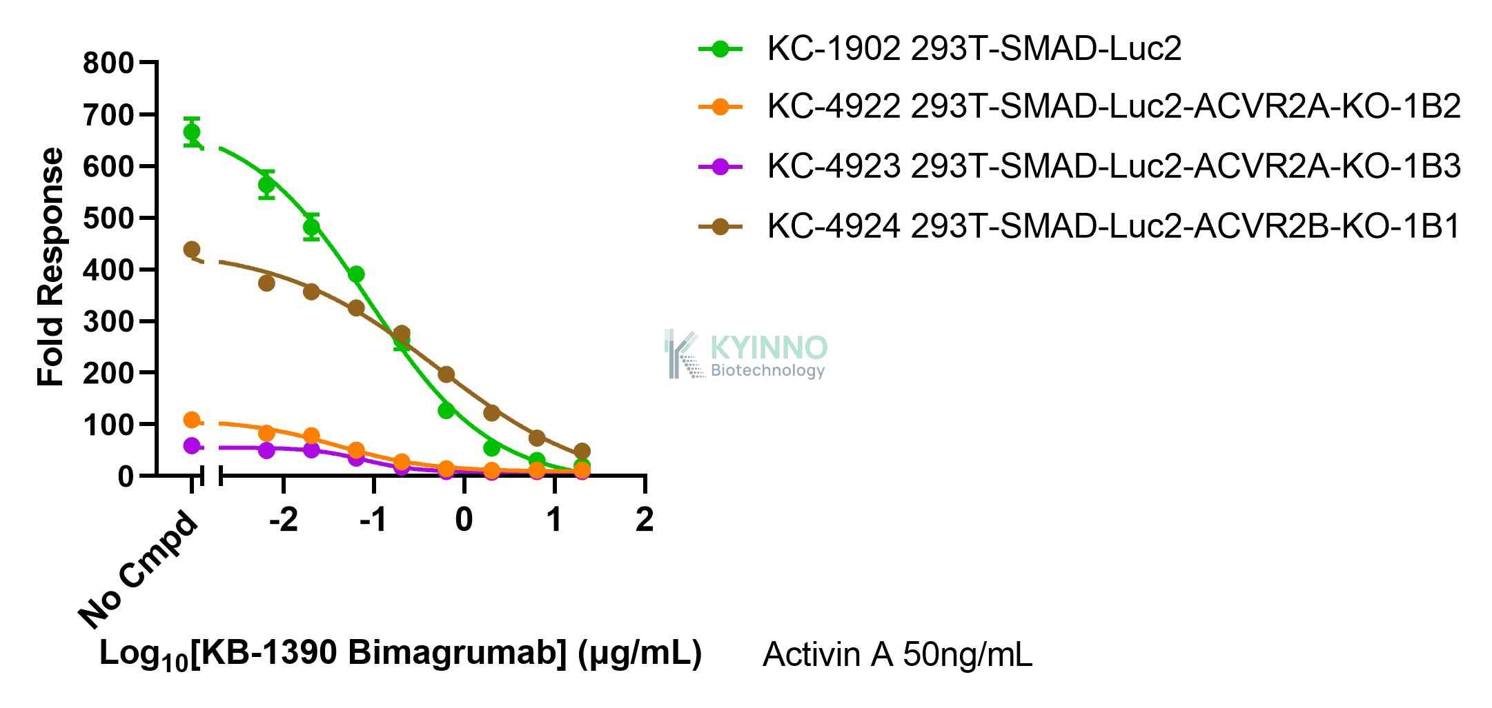
Figure 6. 293T-SMAD-Luc2 cells were seeded into 96-well plates, treated with Bimagrumab (Cat# KB-1390, Kyinno) for 1 hours, and then treated with 50ng/mL Activin A for 16 hours, and then read out using Bright-Glo Detection System.

Cell Resuscitation

1. Prewarm culture medium (DMEM supplemented with 10% FBS, 150μg/ml Hygromycin)in a 37°C water bath.
2. Thaw the frozen vial in a 37°C water bath for 1-2 minutes.
3. Transfer the vial into biosafety cabinet, and wipe the surface with 70% ethanol.
4. Unscrew the top of the vial and transfer the cell suspension gently into a sterile centrifuge tube containing 9.0mL complete culture medium.
5. Spin at ~ 125 × g for 5-7 minutes at room temperature, and discard the supernatant without disturbing the pellet.
6. Resuspend cell pellet with the appropriate volume of complete medium and transfer the cell suspension into a T25 culture flask.
7. Incubate the flask at 37°C, 5% CO2 incubator.
8. Split saturated culture 1:4-1:8 every 2-3 days; seed out at about 1-3 × 10⁵ cells/mL.

Cell Freezing

  1. Prepare the freezing medium (70% DMEM + 20% FBS + 10% DMSO) fresh immediately before use.
  2. Keep the freezing medium on ice and label cryovials.
  3. Transfer cells to a sterile, conical centrifuge tube, and count the cells.
  4. Centrifuge the cells at 250×g for 5 minutes at room temperature and carefully aspirate off the medium.
  5. Resuspend the cells at a density of at least 3×10⁶ cells/mL in chilled freezing medium.
  6. Aliquot 1 mL of the cell suspension into each cryovial.
  7. Freeze cells in the CoolCell freezing container overnight in a -80°C freezer.
  8. Transfer vials to liquid nitrogen for long-term storage

References

  1. Hu HH, Chen DQ, Wang YN, Feng YL, Cao G, Vaziri ND, Zhao YY. New insights into TGF-β/Smad signaling in tissue fibrosis. Chem Biol Interact. 2018 Aug 25;292:76-83. doi: 10.1016/j.cbi.2018.07.008. Epub 2018 Jul 11. PMID: 30017632.
  2. Massagué J, Seoane J, Wotton D. Smad transcription factors. Genes Dev. 2005 Dec 1;19(23):2783-810. doi: 10.1101/gad.1350705. PMID: 16322555.

Use License Agreement

Research Use Only.
Not for use in diagnostic procedures or therapeutic applications.
Redistribution of the cell line or its derivatives is prohibited without prior written permission from Kyinno Biotechnology.
请在浏览器中启用JavaScript来完成此表单。