KC-3563

293T-CLDN1 Cell Line

×
Please enable JavaScript in your browser to complete this form.
26403
Home » 293T-CLDN1 Cell Line

Background of 293T-CLDN1 Cell Line

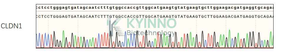
Tight junctions represent one mode of cell-to-cell adhesion in epithelial or endothelial cell sheets, forming continuous seals around cells and serving as a physical barrier to prevent solutes and water from passing freely through the paracellular space. These junctions are comprised of sets of continuous networking strands in the outwardly facing cytoplasmic leaflet, with complementary grooves in the inwardly facing extracytoplasmic leaflet. The protein encoded by this gene, a member of the claudin family, is an integral membrane protein and a component of tight junction strands. Loss of function mutations result in neonatal ichthyosis-sclerosing cholangitis syndrome.

Specifications

Catalog NumberKC-3563
Cell Line Name293T-CLDN1 Cell Line
Clone Number5#
Host Cell Line293T
DescriptionStable 293T cell line expressing exogenous CLDN1 gene
QuantityTwo vials of frozen cells (≥2-106/vial)
StabilityStable in culture over a minimum of 10 passages
ApplicationDrug screening and biological assays
Freezing Medium70% DMEM + 20% FBS + 10% DMSO
Propagation MediumDMEM + 10% FBS + 1μg/mL Puromycin
Selection MarkerPuromycin
MorphologyEpithelial
SubcultureSplit saturated culture 1:4-1:5 every 2-3 days; seed out at about 1-3 × 10⁵ cells/mL
Incubation37 °C with 5% CO2
StorageLiquid nitrogen immediately upon receiving
Doubling TimeApproximately 30 hours
Mycoplasma StatusNegative
In Vivo ValidationNA

Cell Line Generation

293T-CLDN1 cell line was generated using a lentiviral vector expressing the CLDN1 sequence.

Characterization

Figure 1: Characterization of CLDN1 overexpression in the 293T-CLDN1 stable clone using FACS.(Primary antibody: OM-7D3-B3-hIgG1, Cat#KB-1262, Kyinno)

Figure 2: Characterization of CLDN1 overexpressing in 293T-CLDN1 stable clone using PCR sequencing.

Cell Resuscitation

  1. Prewarm culture medium (DMEM supplemented with 10% FBS and 1μg/mL Puromycin)in a 37°C water bath.
  2. Thaw the frozen vial in a 37°C water bath for 1-2 minutes.
  3. Transfer the vial into biosafety cabinet, and wipe the surface with 70% ethanol.
  4. Unscrew the top of the vial and transfer the cell suspension gently into a sterile centrifuge tube containing 9.0mL complete culture medium.
  5. Spin at ~ 125 × g for 5-7 minutes at room temperature, and discard the supernatant without disturbing the pellet.
  6. Resuspend cell pellet with the appropriate volume of complete medium and transfer the cell suspension into a T25 culture flask.
  7. Incubate the flask at 37°C, 5% CO2 incubator.
  8. Split saturated culture 1:4-1:5 every 2-3 days; seed out at about 1-3 × 10⁵ cells/mL.

Cell Freezing

  1. Prepare the freezing medium (70% DMEM + 20% FBS + 10% DMSO) fresh immediately before use.
  2. Keep the freezing medium on ice and label cryovials.
  3. Transfer cells to a sterile, conical centrifuge tube, and count the cells.
  4. Centrifuge the cells at 250×g for 5 minutes at room temperature and carefully aspirate off the medium.
  5. Resuspend the cells at a density of at least 3×10⁶ cells/mL in chilled freezing medium.
  6. Aliquot 1 mL of the cell suspension into each cryovial.
  7. Freeze cells in the CoolCell freezing container overnight in a -80°C freezer.
  8. Transfer vials to liquid nitrogen for long-term storage.

References

  1. Ma F, Ding X, Fan Y, Ying J, Zheng S, Lu N, Xu B. A CLDN1-negative phenotype predicts poor prognosis in triple-negative breast cancer. PLoS One. 2014 Nov 13;9(11):e112765. doi: 10.1371/journal.pone.0112765. PMID: 25393310; PMCID: PMC4231092.
  2. Ouban A, Ahmed A. Analysis of the distribution and expression of claudin-1 tight junction protein in the oral cavity. Appl Immunohistochem Mol Morphol. 2015 Jul;23(6):444-8. doi: 10.1097/PAI.0000000000000104. PMID: 25517868.
Please enable JavaScript in your browser to complete this form.