KC-1458

Jurkat-NFAT-Luc2 Cell Line

×
Please enable JavaScript in your browser to complete this form.
26431
Home » Jurkat-NFAT-Luc2 Cell Line

Background of Jurkat-NFAT-Luc2 Cell Line

NFAT (nuclear factor of activator T cells) proteins belongs to a family of transcription factors that are very important in T cells activation, differentiation and tolerance. NFAT pathway is activated by Ca2+ and the Ca2+/calmodulin dependent serine phosphatase calcineurin after the engagement of T cell receptor (TCR).

Specifications

Catalog NumberKC-1458
Cell Line NameJurkat-NFAT-Luc2 Cell Line
Host Cell LineJurkat
DescriptionJurkat cell line stable expressing exogenous luciferase gene under the control of
NFAT responsive element.
QuantityTwo vials of frozen cells (≥2-106/vial)
StabilityStable in culture over a minimum of 10 passages
ApplicationDrug screening and biological assays
Freezing Medium70% RPMI1640+20% FBS+10% DMSO
Propagation MediumRPMI1640+10% FBS+300ug/ml Hygromycin
Selection MarkerHygromycin B
MorphologyLymphoblast
SubcultureSplit saturated culture 1:4-1:5 every 2-3 days; seed out at about 1-3 × 10⁵ cells/mL
Incubation37 °C with 5% CO2
StorageLiquid nitrogen immediately upon receiving
Doubling TimeApproximately 30 hours
Mycoplasma StatusNegative
In Vivo ValidationNA

Cell Line Generation

Jurkat-NFAT-Luc2 cell line was generated using lentivirus method.

Characterization

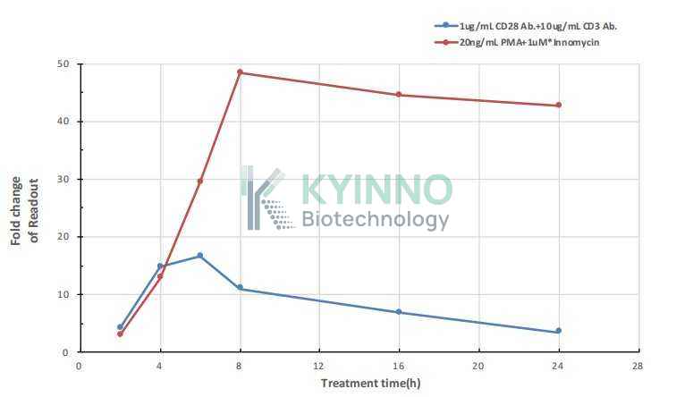
Figure 1. Activation of Jurkat-NFAT-Luc2 cell line stimulated with CD3/CD28 antibodies or PMA and ionomycin. 20,000 Jurkat-NFAT-Luc2 cell line was seeded into the 96-well plate, and treated with differentstimulus, then readout with Bright-Glo system.

Cell Resuscitation

  1. Prewarm culture medium (RPMI1640 supplemented with 10% FBS, 300μg/ml Hygromycin)in a 37°C water bath.
  2. Thaw the frozen vial in a 37°C water bath for 1-2 minutes.
  3. Transfer the vial into biosafety cabinet, and wipe the surface with 70% ethanol.
  4. Unscrew the top of the vial and transfer the cell suspension gently into a sterile centrifuge tube containing 9.0mL complete culture medium.
  5. Spin at ~ 125 × g for 5-7 minutes at room temperature, and discard the supernatant without disturbing the pellet.
  6. Resuspend cell pellet with the appropriate volume of complete medium and transfer the cell suspension into a T25 culture flask.
  7. Incubate the flask at 37°C, 5% CO2 incubator.
  8. Split saturated culture 1:4-1:5 every 2-3 days; seed out at about 1-3 × 10⁵ cells/mL.

Cell Freezing

  1. Prepare the freezing medium (70% RPMI1640 + 20% FBS + 10% DMSO) fresh immediately before use.
  2. Keep the freezing medium on ice and label cryovials.
  3. Transfer cells to a sterile, conical centrifuge tube, and count the cells.
  4. Centrifuge the cells at 250×g for 5 minutes at room temperature and carefully aspirate off the medium.
  5. Resuspend the cells at a density of at least 3×10⁶ cells/mL in chilled freezing medium.
  6. Aliquot 1 mL of the cell suspension into each cryovial.
  7. Freeze cells in the CoolCell freezing container overnight in a -80°C freezer.
  8. Transfer vials to liquid nitrogen for long-term storage.

References

1.Macian, F. NFAT proteins: Key regulators of T-cell development and function. Nat. Rev. Immunol. 5, 472–484 (2005).
2.Hogan, P. G., Chen, L., Nardone, J. & Rao, A. Transcriptional regulation by calcium, calcineurin, and NFAT. Genes Dev. 17, 2205–32 (2003).
Please enable JavaScript in your browser to complete this form.