KC-2953

PC3-Human-FAP-High Cell Line

×
Please enable JavaScript in your browser to complete this form.
26471
Home » PC3-Human-FAP-High Cell Line

Background of PC3-Human-FAP-High Cell Line

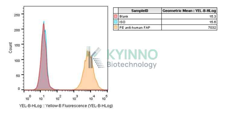
Fibroblast activation protein-α (FAP) is a type II integral serine protease that is specifically expressed by activated fibroblasts. FAP-α displays both exopeptidase and endopeptidase/gelatinase/collagenase activities. FAP-α protein and/or activity have been associated with fibrosis, inflammation and cancer, but the protein is undetectable in most normal tissues. Cancer-associated fibroblasts (CAFs) in the tumor stroma have an abundant and stable expression of FAP, which plays an important role in promoting tumor growth, invasion, metastasis, and immunosuppression. CAF overexpression of FAP promotes tumor development and metastasis by influencing extracellular matrix remodeling, intracellular signaling, angiogenesis, epithelial-to-mesenchymal transition, and immunosuppression.

Specifications

Catalog NumberKC-2953
Cell Line NamePC3-Human-FAP-High Cell Line
Clone NumberNA
Host Cell LinePC3
DescriptionStable PC3 clone expressing exogenous human FAP gene
QuantityTwo vials of frozen cells (≥2-106/vial)
StabilityStable in culture over a minimum of 10 passages
ApplicationDrug screening and biological assays
Freezing Medium70% Ham's F12K + 20% FBS + 10% DMSO
Propagation MediumHam's F12K+10%FBS+1µg/mL Puromycin
Selection MarkerPuromycin
MorphologyEpithelial
SubcultureSplit saturated culture 1:3-1:4 every 2-3 days; seed out at about 1-3 × 10⁵ cells/mL
Incubation37 °C with 5% CO2
StorageLiquid nitrogen immediately upon receiving
Doubling TimeApproximately 35 hours
Mycoplasma StatusNegative
In Vivo ValidationNA

Cell Line Generation

PC3-Human-FAP-High cell line was generated using a lentiviral vector expressing the human FAP sequence.

Characterization

Figure 1: Characterization of human FAP overexpression in the PC3 stable clone using FACS.

Cell Resuscitation

1. Prewarm culture medium (Ham\'s F12K+10%FBS+1µg/mL Puromycin)in a 37°C water bath.
2. Thaw the frozen vial in a 37°C water bath for 1-2 minutes.
3. Transfer the vial into biosafety cabinet, and wipe the surface with 70% ethanol.
4. Unscrew the top of the vial and transfer the cell suspension gently into a sterile centrifuge tube containing 9.0mL complete culture medium.
5. Spin at ~ 125 × g for 5-7 minutes at room temperature, and discard the supernatant without disturbing the pellet.
6. Resuspend cell pellet with the appropriate volume of complete medium and transfer the cell suspension into a T25 culture flask.
7. Incubate the flask at 37°C, 5% CO2 incubator.
8. Split saturated culture 1:3-1:4 every 2-3 days; seed out at about 1-3 × 10⁵ cells/mL.

Cell Freezing

1. Prepare the freezing medium (70% Ham\'s F12K + 20% FBS + 10% DMSO) fresh immediately before use.
2. Keep the freezing medium on ice and label cryovials.
3. Transfer cells to a sterile, conical centrifuge tube, and count the cells.
4. Centrifuge the cells at 250×g for 5 minutes at room temperature and carefully aspirate off the medium.
5. Resuspend the cells at a density of at least 3×10⁶ cells/mL in chilled freezing medium.
6. Aliquot 1 mL of the cell suspension into each cryovial.
7. Freeze cells in the CoolCell freezing container overnight in a -80°C freezer.
8. Transfer vials to liquid nitrogen for long-term storage.

References

1.Juillerat-Jeanneret L, Tafelmeyer P, Golshayan D. Fibroblast activation protein-α in fibrogenic disorders and cancer: more than a prolyl-specific peptidase? Expert Opin Ther Targets. 2017 Oct;21(10):977-991. doi: 10.1080/14728222.2017.1370455. Epub 2017 Aug 30. PMID: 28829211.
2. Xin L, Gao J, Zheng Z, Chen Y, Lv S, Zhao Z, Yu C, Yang X, Zhang R. Fibroblast Activation Protein-α as a Target in the Bench-to-Bedside Diagnosis and Treatment of Tumors: A Narrative Review. Front Oncol. 2021 Aug 19;11:648187. doi:10.3389/fonc.2021.648187. PMID: 34490078; PMCID: PMC8416977.
Please enable JavaScript in your browser to complete this form.