KC-3382

CHOK1-cyno-CD20-Cell-Line

×
Please enable JavaScript in your browser to complete this form.
26979
Home » CHOK1-cyno-CD20-Cell-Line

Background of CHOK1-cyno-CD20-Cell-Line

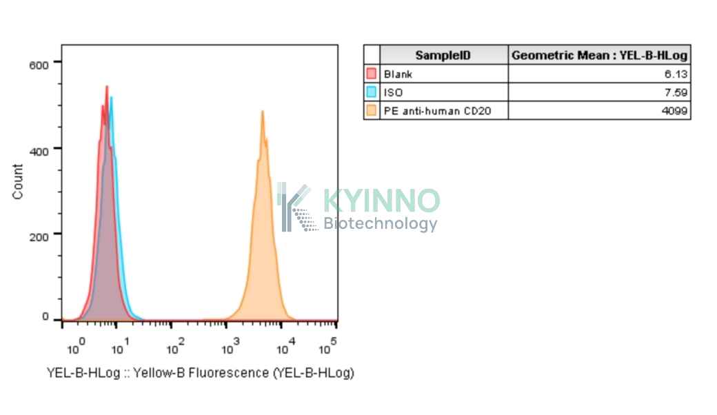
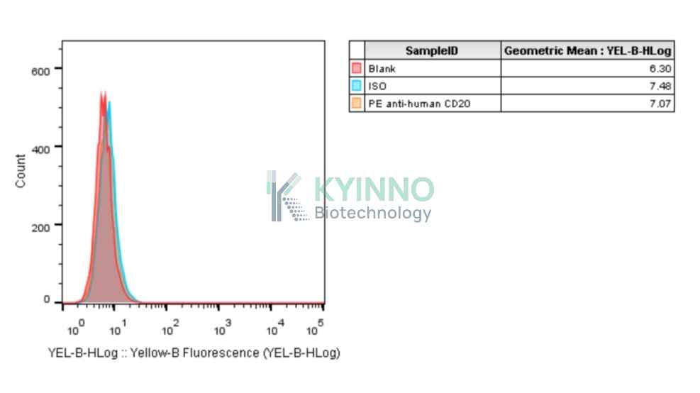
CD20, This gene encodes a member of the membrane-spanning 4A gene family. Members of this nascent protein family are characterized by common structural features and similar intron/exon splice boundaries and display unique expression patterns among hematopoietic cells and nonlymphoid tissues. This gene encodes a B-lymphocyte surface molecule which plays a role in the development and differentiation of B-cells into plasma cells. This family member is localized to 11q12, among a cluster of family members. Alternative splicing of this gene results in two transcript variants which encode the same protein. CD20 is highly expressed in several types of B-cell lymphoma and is an intuitive target for chimeric antigen receptor (CAR) T-cell therapy.

Specifications

Catalog NumberKC-3382
Cell Line NameCHOK1-cyno-CD20-Cell-Line
Host Cell LineCHOK1
DescriptionStable CHOK1 clone expressing exogenous cyno-CD20 gene.
QuantityTwo vials of frozen cells (≥2-106/vial)
StabilityStable in culture over a minimum of 10 passages
ApplicationDrug screening and biological assays
Freezing Medium70% RPMI 1640 + 20% FBS + 10% DMSO
Propagation MediumRPMI 1640 + 10% FBS + 10μg/mL Puromycin
Selection MarkerPuromycin
MorphologyEpithelial
SubcultureSplit saturated culture 1:6-1:8 every 2-3 days; seed out at about 1-3 × 10⁵ cells/mL
Incubation37 °C with 5% CO2
StorageLiquid nitrogen immediately upon receiving
Doubling TimeApproximately 24 hours
Mycoplasma StatusNegative
In Vivo ValidationNA

Cell Line Generation

CHOK1-cyno-CD20-cell-line was generated using a lentiviral vector expressing the cyno-CD20 sequence.

Characterization

Figure 1: Characterization of cyno-CD20 overexpression in the CHOK1-cyno-CD20 stable clone using FACS.

Figure 2: Characterization of cyno-CD20 overexpression in the CHOK1 stable clone using FACS.

Cell Resuscitation

  1. Prewarm culture medium (RPMI 1640 supplemented with 10% FBS and 10μg/mL puromycin)in a 37°C water bath.
  2. Thaw the frozen vial in a 37°C water bath for 1-2 minutes.
  3. Transfer the vial into biosafety cabinet, and wipe the surface with 70% ethanol.
  4. Unscrew the top of the vial and transfer the cell suspension gently into a sterile centrifuge tube containing 9.0mL complete culture medium.
  5. Spin at ~ 125 × g for 5-7 minutes at room temperature, and discard the supernatant without disturbing the pellet.
  6. Resuspend cell pellet with the appropriate volume of complete medium and transfer the cell suspension into a T25 culture flask.
  7. Incubate the flask at 37°C, 5% CO2 incubator.
  8. Split saturated culture 1:6-1:8 every 2-3 days; seed out at about 1-3 × 10⁵ cells/mL.

Cell Freezing

  1. Prepare the freezing medium (70% RPMI 1640 + 20% FBS + 10% DMSO) fresh immediately before use.
  2. Keep the freezing medium on ice and label cryovials.
  3. Transfer cells to a sterile, conical centrifuge tube, and count the cells.
  4. Centrifuge the cells at 250×g for 5 minutes at room temperature and carefully aspirate off the medium.
  5. Resuspend the cells at a density of at least 3×10⁶ cells/mL in chilled freezing medium.
  6. Aliquot 1 mL of the cell suspension into each cryovial.
  7. Freeze cells in the CoolCell freezing container overnight in a -80°C freezer.
  8. Transfer vials to liquid nitrogen for long-term storage.

References

  1. Scheller L, Hudecek M. Engineering CD20 CARs with a Twist. Cancer Immunol Res. 2023 Feb 3;11(2):142-143. doi: 10.1158/2326-6066.CIR-22-0919. PMID: 36633575.
  2. Kim JR, Lee D, Kim Y, Kim JY. CD20/TNFR1 dual-targeting antibody enhances lysosome rupture-mediated cell death in B cell lymphoma. Cancer Immunol Immunother. 2023 Jun;72(6):1567-1580. doi: 10.1007/s00262-022-03344-9. Epub 2022 Dec 19. PMID: 36534148.
Please enable JavaScript in your browser to complete this form.