KC-3416

293T-CD20-Cell-Line

×
Please enable JavaScript in your browser to complete this form.
26991
Home » 293T-CD20-Cell-Line

Background of 293T-CD20-Cell-Line

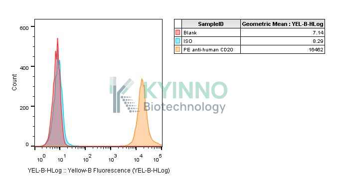
CD20, also known as MS4A1. This gene encodes a member of the membrane-spanning 4A gene family. Members of this nascent protein family are characterized by common structural features and similar intron/exon splice boundaries and display unique expression patterns among hematopoietic cells and nonlymphoid tissues. This gene encodes a B-lymphocyte surface molecule which plays a role in the development and differentiation of B-cells into plasma cells. This family member is localized to 11q12, among a cluster of family members. Alternative splicing of this gene results in two transcript variants which encode the same protein. Research has shown that CD8CD20 T subset functions in host cancer immunosurveillance and tumor microenvironment suppresses this T subset through a PD-L1-dependent mechanism.

Specifications

Catalog NumberKC-3416
Cell Line Name293T-CD20-Cell-Line
Host Cell Line293T
DescriptionStable 293T clone expressing exogenous CD20 gene
QuantityTwo vials of frozen cells (≥2-106/vial)
StabilityStable in culture over a minimum of 10 passages
ApplicationDrug screening and biological assays
Freezing Medium70% DMEM + 20% FBS + 10% DMSO
Propagation MediumDMEM + 10% FBS + 1 μg/mL Puromycin
Selection MarkerPuromycin
MorphologyFibroblastoid
SubcultureSplit saturated culture 1:4-1:5 every 2-3 days; seed out at about 1-3 × 10⁵ cells/mL
Incubation37 °C with 5% CO2
StorageLiquid nitrogen immediately upon receiving
Doubling TimeApproximately 30 hours
Mycoplasma StatusNegative
In Vivo ValidationNA

Cell Line Generation

293T CD20 cell line was generated using a lentiviral vector expressing the CD20 sequence.

Characterization

Figure 1: Characterization of endogenous CD20 expression in 293T cell using FACS.

Figure 2: Characterization of CD20 overexpression in the 293T CD20 stable clone using FACS.

Cell Resuscitation

  1. Prewarm culture medium (DMEM supplemented with 10% FBS and 1 μg/mL puromycin)in a 37°C water bath.
  2. Thaw the frozen vial in a 37°C water bath for 1-2 minutes.
  3. Transfer the vial into biosafety cabinet, and wipe the surface with 70% ethanol.
  4. Unscrew the top of the vial and transfer the cell suspension gently into a sterile centrifuge tube containing 9.0mL complete culture medium.
  5. Spin at ~ 125 × g for 5-7 minutes at room temperature, and discard the supernatant without disturbing the pellet.
  6. Resuspend cell pellet with the appropriate volume of complete medium and transfer the cell suspension into a T25 culture flask.
  7. Incubate the flask at 37°C, 5% CO2 incubator.
  8. Split saturated culture 1:4-1:5 every 2-3 days; seed out at about 1-3 × 10⁵ cells/mL.

Cell Freezing

  1. Prepare the freezing medium (70% DMEM + 20% FBS + 10% DMSO) fresh immediately before use.
  2. Keep the freezing medium on ice and label cryovials.
  3. Transfer cells to a sterile, conical centrifuge tube, and count the cells.
  4. Centrifuge the cells at 250×g for 5 minutes at room temperature and carefully aspirate off the medium.
  5. Resuspend the cells at a density of at least 3×10⁶ cells/mL in chilled freezing medium.
  6. Aliquot 1 mL of the cell suspension into each cryovial.
  7. Freeze cells in the CoolCell freezing container overnight in a -80°C freezer.
  8. Transfer vials to liquid nitrogen for long-term storage

References

  1. Mudd TW Jr, Lu C, Klement JD, Liu K. MS4A1 expression and function in T cells in the colorectal cancer tumor microenvironment. Cell Immunol. 2021 Feb;360:104260. doi: 10.1016/j.cellimm.2020.104260. Epub 2020 Dec 14. PMID: 33352466; PMCID: PMC7855947.
Please enable JavaScript in your browser to complete this form.