KC-3862

K562-HLA-A1101-Luc2 Cell Line

×
Please enable JavaScript in your browser to complete this form.
34002
Home » 细胞系 » K562-HLA-A1101-Luc2 Cell Line

Background of K562-HLA-A1101-Luc2 Cell Line

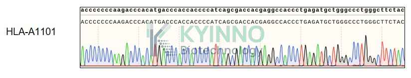
HLA-A belongs to the HLA class I heavy chain paralogues. This class I molecule is a heterodimer consisting of a heavy chain and a light chain (beta-2 microglobulin). The heavy chain is anchored in the membrane. Class I molecules play a central role in the immune system by presenting peptides derived from the endoplasmic reticulum lumen so that they can be recognized by cytotoxic T cells. They are expressed in nearly all cells. The heavy chain is approximately 45 kDa and its gene contains 8 exons. Exon 1 encodes the leader peptide, exons 2 and 3 encode the alpha1 and alpha2 domains, which both bind the peptide, exon 4 encodes the alpha3 domain, exon 5 encodes the transmembrane region, and exons 6 and 7 encode the cytoplasmic tail. Polymorphisms within exon 2 and exon 3 are responsible for the peptide binding specificity of each class one molecule. Typing for these polymorphisms is routinely done for bone marrow and kidney transplantation. More than 6000 HLA-A alleles have been described. The HLA system plays an important role in the occurrence and outcome of infectious diseases, including those caused by the malaria parasite, the human immunodeficiency virus (HIV), and the severe acute respiratory syndrome coronavirus (SARS-CoV). The structural spike and the nucleocapsid proteins of the novel coronavirus SARS-CoV-2, which causes coronavirus disease 2019 (COVID-19), are reported to contain multiple Class I epitopes with predicted HLA restrictions. Individual HLA genetic variation may help explain different immune responses to a virus across a population.

Specifications

Catalog NumberKC-3862
Cell Line NameK562-HLA-A1101-Luc2 Cell Line
Clone Number1#
Host Cell LineK562 Luc2 Cell Line
DescriptionStable K562 HLA-A1101 Luc2 cell line expressing exogenous Luc2 gene
QuantityTwo vials of frozen cells (≥2-106/vial)
StabilityStable in culture over a minimum of 10 passages
ApplicationDrug screening and biological assays
Freezing Medium70% RPMI1640+20% FBS+10% DMSO
Propagation MediumRPMI1640 + 10% FBS + 1ug/ml Puromycin + 300ug/ml HygromycinB
Selection MarkerPuromycin, Hygromycin B
MorphologyMostly single, round (some polymorph) cells in suspension
SubcultureSplit saturated culture 1:2-1:4 every 2-3 days; seed out at about 1-3 × 10⁵ cells/mL
Incubation37 °C with 5% CO2
StorageLiquid nitrogen immediately upon receiving
Doubling TimeApproximately 30 hours
Mycoplasma StatusNegative
In Vivo ValidationNA

Cell Line Generation

K562 HLA-A1101 Luc2 Cell Line was generated using a lentiviral vector expressing the HLA-A1101 sequence.

Characterization

Figure 1: Characterization of HLA-A1101 overexpression in K562 HLA-A1101 Luc2 stable clone using FACS.

Figure2: Characterization of HLA-A0101 overexpressing in K562 HLA-A1101 Luc2 stable clone using PCR sequencing.

Cell Resuscitation

  1. Prewarm culture medium (RPMI1640 supplemented with 10% FBS and 1ug/ml puromycin + 300ug/ml HygromycinB)in a 37°C water bath.
  2. Thaw the frozen vial in a 37°C water bath for 1-2 minutes.
  3. Transfer the vial into biosafety cabinet, and wipe the surface with 70% ethanol.
  4. Unscrew the top of the vial and transfer the cell suspension gently into a sterile centrifuge tube containing 9.0mL complete culture medium.
  5. Spin at ~ 125 × g for 5-7 minutes at room temperature, and discard the supernatant without disturbing the pellet.
  6. Resuspend cell pellet with the appropriate volume of complete medium and transfer the cell suspension into a T25 culture flask.
  7. Incubate the flask at 37°C, 5% CO2 incubator.
  8. Split saturated culture 1:2-1:4 every 2-3 days; seed out at about 1-3 × 10⁵ cells/mL.

Cell Freezing

  1. Prepare the freezing medium (70% RPMI1640 + 20% FBS + 10% DMSO) fresh immediately before use.
  2. Keep the freezing medium on ice and label cryovials.
  3. Transfer cells to a sterile, conical centrifuge tube, and count the cells.
  4. Centrifuge the cells at 250×g for 5 minutes at room temperature and carefully aspirate off the medium.
  5. Resuspend the cells at a density of at least 3×10⁶ cells/mL in chilled freezing medium.
  6. Aliquot 1 mL of the cell suspension into each cryovial.
  7. Freeze cells in the CoolCell freezing container overnight in a -80°C freezer.
  8. Transfer vials to liquid nitrogen for long-term storage.

References

  1. Lin CL, Lo WF, Lee TH, Ren Y, Hwang SL, Cheng YF, Chen CL, Chang YS, Lee SP, Rickinson AB, Tam PK. Immunization with Epstein-Barr Virus (EBV) peptide-pulsed dendritic cells induces functional CD8+ T-cell immunity and may lead to tumor regression in patients with EBV-positive nasopharyngeal carcinoma. Cancer Res. 2002 Dec 1;62(23):6952-8. PMID: 12460912.
  2. 2.Di D, Nunes JM, Jiang W, Sanchez-Mazas A. Like Wings of a Bird: Functional Divergence and Complementarity between HLA-A and HLA-B Molecules. Mol Biol Evol. 2021 Apr 13;38(4):1580-1594. doi: 10.1093/molbev/msaa325. PMID: 33320202; PMCID: PMC8355449.
Please enable JavaScript in your browser to complete this form.