KC-3139

293T-CRE-Luc2-GCGR-Cell-Line

×
Please enable JavaScript in your browser to complete this form.
34162
Home » 293T-CRE-Luc2-GCGR-Cell-Line

Background of 293T-CRE-Luc2-GCGR-Cell-Line

"Cyclic adenosine monophosphate (cAMP), the first discovered second messenger, plays a key role in cell signaling, regulating many physiological and pathological processes. cAMP can regulate the transcription of a variety of target genes, primarily through protein kinase A (PKA) and its downstream effectors such as cAMP-responsive element binding protein (CREB).
Abnormal elevations of glucagon (GCG) are the leading cause of type II diabetes. When GCG interacts with glucagon receptor (GCGR), GCG can increase blood sugar levels."

Specifications

Catalog NumberKC-3139
Cell Line Name293T-CRE-Luc2-GCGR-Cell-Line
Host Cell Line293T-CRE-Luc2
DescriptionStable 293T-CRE-Luc2 cell line expressing exogenous GCGR gene
QuantityTwo vials of frozen cells (≥2-106/vial)
StabilityStable in culture over a minimum of 10 passages
ApplicationDrug screening and biological assays
Freezing Medium70% DMEM+20% FBS+10% DMSO
Propagation MediumDMEM+10% FBS +150μg/mL Hygromycin+1μg/mL Puromycin
Selection MarkerHygromycin and Puromycin
MorphologyEpithelial
SubcultureSplit saturated culture 1:4-1:5 every 2-3 days; seed out at about 1-3 × 10⁵ cells/mL
Incubation37 °C with 5% CO2
StorageLiquid nitrogen immediately upon receiving
Doubling TimeApproximately 30 hours
Mycoplasma StatusNegative
In Vivo ValidationNA

Cell Line Generation

293T-CRE-Luc2-GCGR cell line was generated using lentivirus expressing GCGR sequence.

Characterization

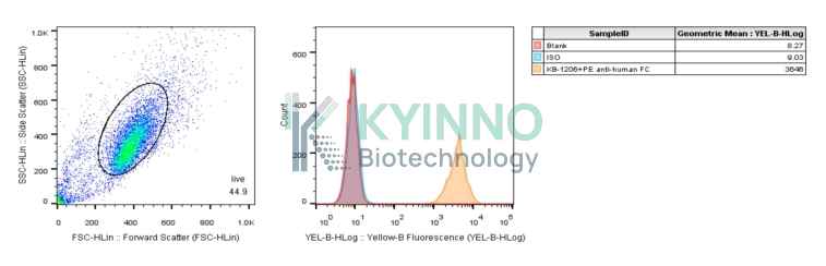
Figure 1: Characterization of human GCGR overexpression in 293T-CRE-Luc2 stable clones using FCAS.

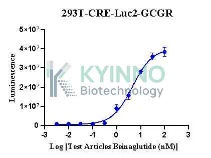
Figure 2: 293T-CRE-Luc2-GCGR cells were seeded into the 96-well plate, treated with GCG 6 hours, then readout with Bright-lite™ Luciferase Assay system.

Cell Resuscitation

  1. Prewarm culture medium (DMEM supplemented with 10% FBS, 150μg/mL Hygromycin and 1μg/mL Puromycin)in a 37°C water bath.
  2. Thaw the frozen vial in a 37°C water bath for 1-2 minutes.
  3. Transfer the vial into biosafety cabinet, and wipe the surface with 70% ethanol.
  4. Unscrew the top of the vial and transfer the cell suspension gently into a sterile centrifuge tube containing 9.0mL complete culture medium.
  5. Spin at ~ 125 × g for 5-7 minutes at room temperature, and discard the supernatant without disturbing the pellet.
  6. Resuspend cell pellet with the appropriate volume of complete medium and transfer the cell suspension into a T25 culture flask.
  7. Incubate the flask at 37°C, 5% CO2 incubator.
  8. Split saturated culture 1:4-1:8 every 2-3 days; seed out at about 1-3 × 10⁵ cells/mL.

Cell Freezing

  1. Prepare the freezing medium (70% DMEM + 20% FBS + 10% DMSO) fresh immediately before use.
  2. Keep the freezing medium on ice and label cryovials.
  3. Transfer cells to a sterile, conical centrifuge tube, and count the cells.
  4. Centrifuge the cells at 250×g for 5 minutes at room temperature and carefully aspirate off the medium.
  5. Resuspend the cells at a density of at least 3×10⁶ cells/mL in chilled freezing medium.
  6. Aliquot 1 mL of the cell suspension into each cryovial.
  7. Freeze cells in the CoolCell freezing container overnight in a -80°C freezer.
  8. Transfer vials to liquid nitrogen for long-term storage

References

  1. Sands WA, Palmer TM. Regulating gene transcription in response to cyclic AMP elevation. Cell Signal. 2008 Mar;20(3):460-6. doi: 10.1016/j.cellsig.2007.10.005. Epub 2007 Oct 12. PMID: 17993258.
  2. Verleye L, Ottevanger PB, van der Graaf W, Reed NS, Vergote I; Gynaecological Cancer Group (GCG) of European Organisation for Research and Treatment of Cancer (EORTC). EORTC-GCG process quality indicators for ovarian cancer surgery. Eur J Cancer. 2009 Mar;45(4):517-26. doi: 10.1016/j.ejca.2008.09.031. Epub 2008 Nov 14. PMID: 19013789.
Please enable JavaScript in your browser to complete this form.