KC-3327

MC38-cMet Cell Line

Stable MC38 cell line expressing exogenous human c-Met gene

×
Please enable JavaScript in your browser to complete this form.
34318
Home » MC38-cMet Cell Line

Background of MC38-cMet Cell Line

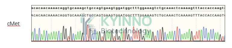
c-Met, also named as hepatocyte growth factor receptor (HGFR), is a single pass tyrosine kinase receptor, which is mainly expressed on the cells of epithelial origin, and play essential role in embryonic development, organogenesis and wound healing. HGF and its splicing isoform are the only know ligands of c-Met. Abnormal activation of c-Met due to overexpression of Met or its ligands, fusion mutation as well as exon14 skipping have been implicated In oncogenesis.

Specifications

Catalog NumberKC-3327
Cell Line NameMC38-cMet Cell Line
NCBI/UniProt Accession NumberNM_001127500.3
Clone Number1#
Host Cell LineMC38
DescriptionStable MC38 cell line expressing exogenous human MET gene
QuantityTwo vials of frozen cells (≥2-106/vial)
StabilityStable in culture over a minimum of 10 passages
ApplicationDrug screening and biological assays
Freezing Medium70% DMEM + 20% FBS + 10% DMSO
Propagation MediumDMEM + 10% FBS + 5μg/mL Puromycin
Selection MarkerPuromycin
MorphologyEpithelial
SubcultureSplit saturated culture 1:4-1:8 every 2-3 days; seed out at about 1-3 × 10⁵ cells/mL
Incubation37 °C with 5% CO2
StorageLiquid nitrogen immediately upon receiving
Doubling TimeApproximately 24 hours
Mycoplasma StatusNegative
In Vivo ValidationNA

Cell Line Generation

MC38-cMet cell line was generated using a lentiviral vector expressing the human MET sequence.

Characterization

Figure 1: Characterization of human MET overexpression in MC38-cMet stable clones using FACS.

Figure 2: Characterization of MC38-cMet Cell Line stable clone using PCR sequencing.

Cell Resuscitation

1. Prewarm culture medium (DMEM supplemented with 10% FBS and 5μg/mL puromycin)in a 37°C water bath.
2. Thaw the frozen vial in a 37°C water bath for 1-2 minutes.
3. Transfer the vial into biosafety cabinet, and wipe the surface with 70% ethanol.
4. Unscrew the top of the vial and transfer the cell suspension gently into a sterile centrifuge tube containing 9.0mL complete culture medium.
5. Spin at ~ 125 × g for 5-7 minutes at room temperature, and discard the supernatant without disturbing the pellet.
6. Resuspend cell pellet with the appropriate volume of complete medium and transfer the cell suspension into a T25 culture flask.
7. Incubate the flask at 37°C, 5% CO2 incubator.
8. Split saturated culture 1:4-1:8 every 2-3 days; seed out at about 1-3 × 10⁵ cells/mL.

Cell Freezing

1. Prepare the freezing medium (70% DMEM + 20% FBS + 10% DMSO) fresh immediately before use.
2. Keep the freezing medium on ice and label cryovials.
3. Transfer cells to a sterile, conical centrifuge tube, and count the cells.
4. Centrifuge the cells at 250×g for 5 minutes at room temperature and carefully aspirate off the medium.
5. Resuspend the cells at a density of at least 3×10⁶ cells/mL in chilled freezing medium.
6. Aliquot 1 mL of the cell suspension into each cryovial.
7. Freeze cells in the CoolCell freezing container overnight in a -80°C freezer.
8. Transfer vials to liquid nitrogen for long-term storage.

References

1. Saffroy, R. et al. MET exon 14 mutations as targets in routine molecular analysis of primary sarcomatoid carcinoma of the lung. Oncotarget 8, 42428-42437 (2017).
2. Peschard, P. & Park, M. From Tpr-Met to Met, tumorigenesi.s and tubes. Oncogene 26, 127&-1285 (2007).
Please enable JavaScript in your browser to complete this form.