KC-3549

CHOK1-mouse-CD137 Cell Line

×
Please enable JavaScript in your browser to complete this form.
34490
Home » CHOK1-mouse-CD137 Cell Line

Background of CHOK1-mouse-CD137 Cell Line

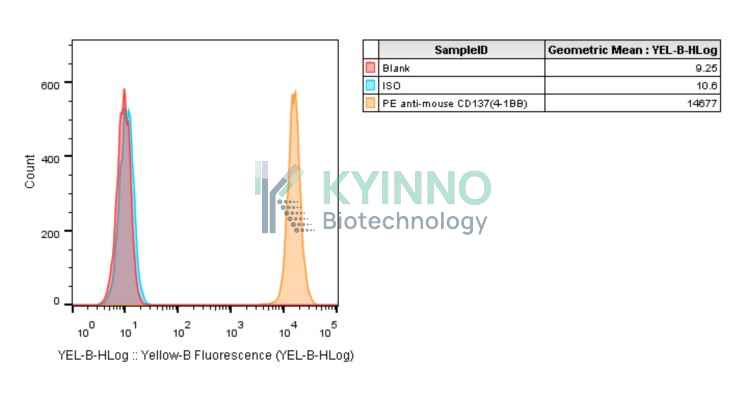
The protein encoded by this gene is a member of the TNF-receptor superfamily. This receptor contributes to the clonal expansion, survival, and development of T cells. It can also induce proliferation in peripheral monocytes, enhance T cell apoptosis induced by TCR/CD3 triggered activation, and regulate CD28 co-stimulation to promote Th1 cell responses. The expression of this receptor is induced by lymphocyte activation. TRAF adaptor proteins have been shown to bind to this receptor and transduce the signals leading to activation of NF-kappaB.

Specifications

Catalog NumberKC-3549
Cell Line NameCHOK1-mouse-CD137 Cell Line
Clone Number1#
Host Cell LineCHOK1
DescriptionStable CHOK1 cell line expressing exogenous mouse-CD137 gene
QuantityTwo vials of frozen cells (≥2-106/vial)
StabilityStable in culture over a minimum of 10 passages
ApplicationDrug screening and biological assays
Freezing Medium70% RPMI1640 + 20% FBS + 10% DMSO
Propagation MediumRPMI1640 + 10% FBS + 10μg/mL Puromycin
Selection MarkerPuromycin
MorphologyEpithelial
SubcultureSplit saturated culture 1:4-1:8 every 2-3 days; seed out at about 1-3 × 10⁵ cells/mL
Incubation37 °C with 5% CO2
StorageLiquid nitrogen immediately upon receiving
Doubling TimeApproximately 30 hours
Mycoplasma StatusNegative
In Vivo ValidationNA

Cell Line Generation

CHOK1-mouse-CD137 cell line was generated using a lentiviral vector expressing the mouse-CD137 sequence.

Characterization

Figure 1: Characterization of mouse-CD137 overexpressing in CHOK1-mouse-CD137 stable clone using FACS.

Figure 2: Characterization of mouse-CD137 overexpressing in CHOK1-mouse-CD137 stable clone using PCR sequencing.

Cell Resuscitation

  1. Prewarm culture medium (RPMI1640 supplemented with 10% FBS and 10μg/mL Puromycin)in a 37°C water bath.
  2. Thaw the frozen vial in a 37°C water bath for 1-2 minutes.
  3. Transfer the vial into biosafety cabinet, and wipe the surface with 70% ethanol.
  4. Unscrew the top of the vial and transfer the cell suspension gently into a sterile centrifuge tube containing 9.0mL complete culture medium.
  5. Spin at ~ 125 × g for 5-7 minutes at room temperature, and discard the supernatant without disturbing the pellet.
  6. Resuspend cell pellet with the appropriate volume of complete medium and transfer the cell suspension into a T25 culture flask.
  7. Incubate the flask at 37°C, 5% CO2 incubator.
  8. Split saturated culture 1:4-1:8 every 2-3 days; seed out at about 1-3 × 10⁵ cells/mL.

Cell Freezing

  1. Prepare the freezing medium (70% RPMI1640 + 20% FBS + 10% DMSO) fresh immediately before use.
  2. Keep the freezing medium on ice and label cryovials.
  3. Transfer cells to a sterile, conical centrifuge tube, and count the cells.
  4. Centrifuge the cells at 250×g for 5 minutes at room temperature and carefully aspirate off the medium.
  5. Resuspend the cells at a density of at least 3×10⁶ cells/mL in chilled freezing medium.
  6. Aliquot 1 mL of the cell suspension into each cryovial.
  7. Freeze cells in the CoolCell freezing container overnight in a -80°C freezer.
  8. Transfer vials to liquid nitrogen for long-term storage.

References

  1. Söderström LÅ, Gertow K, Folkersen L, Sabater-Lleal M, Sundman E, Sheikine Y, Goel A, Baldassarre D, Humphries SE, de Faire U, Watkins H, Tremoli E, Veglia F, Hamsten A, Hansson GK, Olofsson PS. Human genetic evidence for involvement of CD137 in atherosclerosis. Mol Med. 2014 Oct 14;20(1):456-65. doi: 10.2119/molmed.2014.00004. PMID: 25032953; PMCID: PMC4212009.
  2. He Y, Ao DH, Li XQ, Zhong SS, A R, Wang YY, Xiang YJ, Xu BL, Yang TT, Gao XG, Liu GZ. Increased Soluble CD137 Levels and CD4+ T-Cell-Associated Expression of CD137 in Acute Atherothrombotic Stroke. Clin Transl Sci. 2018 Jul;11(4):428-434. doi: 10.1111/cts.12553. Epub 2018 Apr 26. PMID: 29697202; PMCID: PMC6039206.
Please enable JavaScript in your browser to complete this form.