KC-3688

293T-CDH2 Cell Line

×
请在浏览器中启用JavaScript来完成此表单。
34616
Home » 293T-CDH2 Cell Line

Background of 293T-CDH2 Cell Line

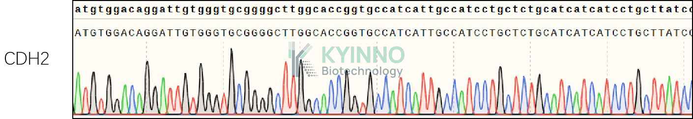
This gene encodes a classical cadherin and member of the cadherin superfamily. Alternative splicing results in multiple transcript variants, at least one of which encodes a preproprotein is proteolytically processed to generate a calcium-dependent cell adhesion molecule and glycoprotein. This protein plays a role in the establishment of left-right asymmetry, development of the nervous system and the formation of cartilage and bone.

Specifications

Catalog NumberKC-3688
Cell Line Name293T-CDH2 Cell Line
NCBI/UniProt Accession NumberNM_001792
Clone Number3#
Host Cell Line293T
DescriptionStable 293T cell line expressing exogenous CDH2 gene
QuantityOne vial of frozen cells (≥2-106/vial)
StabilityStable in culture over a minimum of 10 passages
ApplicationDrug screening and biological assays
Freezing Medium70% DMEM + 20% FBS + 10% DMSO
Propagation MediumDMEM + 10% FBS + 1μg/mL Puromycin
Selection MarkerPuromycin
MorphologyEpithelial
SubcultureSplit saturated culture 1:4-1:5 every 2-3 days; seed out at about 1-3 × 10⁵ cells/mL
Incubation37 °C with 5% CO2
StorageLiquid nitrogen immediately upon receiving
Doubling TimeApproximately 30 hours
Mycoplasma StatusNegative
In Vivo ValidationNA

Cell Line Generation

293T-CDH2 cell line was generated using a lentiviral vector expressing the CDH2 sequence.

Characterization

Figure 1: Characterization of CDH2 overexpression in the 293T-CDH2 stable clone using FACS.

Figure 2: Characterization of CDH2 overexpressing in 293T-CDH2 stable clone using PCR sequencing.

Cell Resuscitation

  1. Prewarm culture medium (DMEM supplemented with 10% FBS and 1μg/mL Puromycin)in a 37°C water bath.
  2. Thaw the frozen vial in a 37°C water bath for 1-2 minutes.
  3. Transfer the vial into biosafety cabinet, and wipe the surface with 70% ethanol.
  4. Unscrew the top of the vial and transfer the cell suspension gently into a sterile centrifuge tube containing 9.0mL complete culture medium.
  5. Spin at ~ 125 × g for 5-7 minutes at room temperature, and discard the supernatant without disturbing the pellet.
  6. Resuspend cell pellet with the appropriate volume of complete medium and transfer the cell suspension into a T25 culture flask.
  7. Incubate the flask at 37°C, 5% CO2 incubator.
  8. Split saturated culture 1:4-1:5 every 2-3 days; seed out at about 1-3 × 10⁵ cells/mL.

Cell Freezing

  1. Prepare the freezing medium (70% DMEM + 20% FBS + 10% DMSO) fresh immediately before use.
  2. Keep the freezing medium on ice and label cryovials.
  3. Transfer cells to a sterile, conical centrifuge tube, and count the cells.
  4. Centrifuge the cells at 250×g for 5 minutes at room temperature and carefully aspirate off the medium.
  5. Resuspend the cells at a density of at least 3×10⁶ cells/mL in chilled freezing medium.
  6. Aliquot 1 mL of the cell suspension into each cryovial.
  7. Freeze cells in the CoolCell freezing container overnight in a -80°C freezer.
  8. Transfer vials to liquid nitrogen for long-term storage.

References

  1. Chung H, Jung H, Jho EH, Multhaupt HAB, Couchman JR, Oh ES. Keratinocytes negatively regulate the N-cadherin levels of melanoma cells via contact-mediated calcium regulation. Biochem Biophys Res Commun. 2018 Sep 5;503(2):615-620. doi: 10.1016/j.bbrc.2018.06.050. Epub 2018 Jun 14. PMID: 29902459.
  2. He K, Yu P, Xing HY, Li Y, Tian Z, Wang M, Tang KJ, Rao Q. [Resistance of leukemia KG1a cells with positive N-cadherin in phase G(0) against killing activity of VP16]. Zhongguo Shi Yan Xue Ye Xue Za Zhi. 2011 Oct;19(5):1102-6. Chinese. PMID: 22040951.

Use License Agreement

Research Use Only.
Not for use in diagnostic procedures or therapeutic applications.
Redistribution of the cell line or its derivatives is prohibited without prior written permission from Kyinno Biotechnology.
请在浏览器中启用JavaScript来完成此表单。