KC-3712

CHOK1-CDH2-Cell-Line

×
Please enable JavaScript in your browser to complete this form.
34644
Home » CHOK1-CDH2-Cell-Line

Background of CHOK1-CDH2-Cell-Line

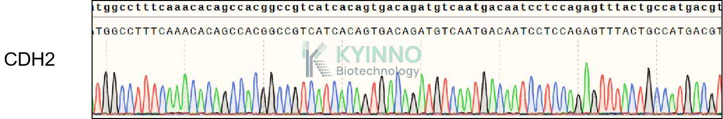
CDH2, also known as CD325. This gene encodes a classical cadherin and member of the cadherin superfamily. Alternative splicing results in multiple transcript variants, at least one of which encodes a preproprotein is proteolytically processed to generate a calcium-dependent cell adhesion molecule and glycoprotein. This protein plays a role in the establishment of left-right asymmetry, development of the nervous system and the formation of cartilage and bone. The expression of N-cadherin increases on the intercellular borders of BM-MSCs through the TGF-β canonical signaling and they collectively migrate in response to breast tumor cells expressing TGF-β via N-cadherin-dependent cell-cell adhesion.

Specifications

Catalog NumberKC-3712
Cell Line NameCHOK1-CDH2-Cell-Line
Host Cell LineCHOK1
DescriptionStable CHOK1 cell line expressing exogenous CDH2 gene
QuantityTwo vials of frozen cells (≥2-106/vial)
StabilityStable in culture over a minimum of 10 passages
ApplicationDrug screening and biological assays
Freezing Medium70% RPMI1640 + 20% FBS + 10% DMSO
Propagation MediumRPMI1640 + 10% FBS + 10μg/ml Puromycin
Selection MarkerPuromycin
MorphologyEpithelial
SubcultureSplit saturated culture 1:4-1:8 every 2-3 days; seed out at about 1-3 × 10⁵ cells/ml
Incubation37 °C with 5% CO2
StorageLiquid nitrogen immediately upon receiving
Doubling TimeApproximately 30 hours
Mycoplasma StatusNegative
In Vivo ValidationNA

Cell Line Generation

CHOK1 CDH2 Cell Line was generated using a lentiviral vector expressing the CDH2 sequence.

Characterization

Figure 1: Characterization of CDH2 overexpression in the CHOK1 CDH2 stable clone using FACS.

Figure 2: Characterization of CDH2 overexpression in CHOK1 stable clone using DNA sequencing.

Cell Resuscitation

  1. Prewarm culture medium (RPMI1640 supplemented with 10% FBS and 10μg/mL Puromycin)in a 37°C water bath.
  2. Thaw the frozen vial in a 37°C water bath for 1-2 minutes.
  3. Transfer the vial into biosafety cabinet, and wipe the surface with 70% ethanol.
  4. Unscrew the top of the vial and transfer the cell suspension gently into a sterile centrifuge tube containing 9.0mL complete culture medium.
  5. Spin at ~ 125 × g for 5-7 minutes at room temperature, and discard the supernatant without disturbing the pellet.
  6. Resuspend cell pellet with the appropriate volume of complete medium and transfer the cell suspension into a T25 culture flask.
  7. Incubate the flask at 37°C, 5% CO2 incubator.
  8. Split saturated culture 1:4-1:8 every 2-3 days; seed out at about 1-3 × 10⁵ cells/mL.

Cell Freezing

  1. Prepare the freezing medium (70% RPMI1640 + 20% FBS + 10% DMSO) fresh immediately before use.
  2. Keep the freezing medium on ice and label cryovials.
  3. Transfer cells to a sterile, conical centrifuge tube, and count the cells.
  4. Centrifuge the cells at 250×g for 5 minutes at room temperature and carefully aspirate off the medium.
  5. Resuspend the cells at a density of at least 3×10⁶ cells/mL in chilled freezing medium.
  6. Aliquot 1 mL of the cell suspension into each cryovial.
  7. Freeze cells in the CoolCell freezing container overnight in a -80°C freezer.
  8. Transfer vials to liquid nitrogen for long-term storage

References

  1. Choi S, Yu J, Kim W, Park KS. N-cadherin mediates the migration of bone marrow-derived mesenchymal stem cells toward breast tumor cells. Theranostics. 2021 May 3;11(14):6786-6799. doi: 10.7150/thno.59703. PMID: 34093853; PMCID: PMC8171089.
Please enable JavaScript in your browser to complete this form.