KC-3785

K562-41BBL-mem-IL21 Cell Line

×
Please enable JavaScript in your browser to complete this form.
34772
Home » 细胞系 » K562-41BBL-mem-IL21 Cell Line

Background of K562-41BBL-mem-IL21 Cell Line

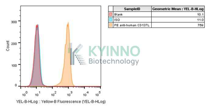
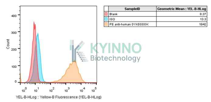
The protein encoded by this gene is a cytokine that belongs to the tumor necrosis factor (TNF) ligand family. This transmembrane cytokine is a bidirectional signal transducer that acts as a ligand for TNFRSF9/4-1BB, which is a costimulatory receptor molecule in T lymphocytes. This cytokine and its receptor are involved in the antigen presentation process and in the generation of cytotoxic T cells. The receptor TNFRSF9/4-1BB is absent from resting T lymphocytes but rapidly expressed upon antigenic stimulation. The ligand encoded by this gene, TNFSF9/4-1BBL, has been shown to reactivate anergic T lymphocytes in addition to promoting T lymphocyte proliferation. This cytokine has also been shown to be required for the optimal CD8 responses in CD8 T cells. This cytokine is expressed in carcinoma cell lines, and is thought to be involved in T cell-tumor cell interaction.
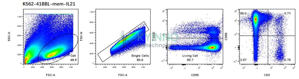
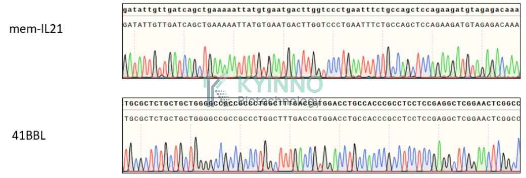
IL21 gene encodes a member of the common-gamma chain family of cytokines with immunoregulatory activity. The encoded protein plays a role in both the innate and adaptive immune responses by inducing the differentiation, proliferation and activity of multiple target cells including macrophages, natural killer cells, B cells and cytotoxic T cells. Dysregulation of this gene plays a role in multiple immune-mediated diseases including lupus, psoriasis and chronic inflammatory diseases. Alternatively spliced transcript variants encoding multiple isoforms have been observed for this gene. [provided by RefSeq, Dec 2011]

Specifications

Catalog NumberKC-3785
Cell Line NameK562-41BBL-mem-IL21 Cell Line
NCBI/UniProt Accession NumberNM_003811.4 & NM_021803.4
Clone NumberNA
Host Cell LineK562
DescriptionStable K562 cell line expressing exogenous 41BBL and mem-IL21 gene
QuantityTwo vials of frozen cells (≥2-106/vial)
StabilityStable in culture over a minimum of 10 passages
ApplicationDrug screening and biological assays
Freezing Medium70% RPMI1640+20% FBS+10% DMSO
Propagation MediumRPMI1640+10%FBS+1µg/mL Puromycin
Selection MarkerPuromycin
MorphologyLymphoblast
SubcultureSplit saturated culture 1:4-1:5 every 2-3 days; seed out at about 1-3 × 10⁵ cells/mL
Incubation37 °C with 5% CO2
StorageLiquid nitrogen immediately upon receiving
Doubling TimeApproximately 30-40 hours
Mycoplasma StatusNegative
In Vivo ValidationNA

Cell Line Generation

K562-41BBL-mem-IL21 cell line was generated using lentivirus expressing 41BBL-mem-IL21 sequence.

Characterization

Figure 1. Characterization of 41BBL over-expression in the K562-41BBL-mem-IL21 stable clone using FACS.

Figure 2. Characterization of Flag (mem-IL21) over-expression in the K562-41BBL-mem-IL21 stable clone using FACS.

Figure 3. In vitro expansion and phenotypic analysis of NK cells supported by K562-41BB6-mem-IL21 feeder cells.

Figure 4. Characterization of mem-IL21 and 41BBL over-expression in the K562-41BBL-mem-IL21 stable clone using PCR.

Cell Resuscitation

  1. Prewarm culture medium (RPMI1640 supplemented with 10% FBS and 1μg/mL Puromycin)in a 37°C water bath.
  2. Thaw the frozen vial in a 37°C water bath for 1-2 minutes.
  3. Transfer the vial into biosafety cabinet, and wipe the surface with 70% ethanol.
  4. Unscrew the top of the vial and transfer the cell suspension gently into a sterile centrifuge tube containing 9.0mL complete culture medium.
  5. Spin at ~ 125 × g for 5-7 minutes at room temperature, and discard the supernatant without disturbing the pellet.
  6. Resuspend cell pellet with the appropriate volume of complete medium and transfer the cell suspension into a T25 culture flask.
  7. Incubate the flask at 37°C, 5% CO2 incubator.
  8. Split saturated culture 1:4-1:5 every 2-3 days; seed out at about 1-3 × 10⁵ cells/mL.

Cell Freezing

  1. Prepare the freezing medium (70% RPMI1640 + 20% FBS + 10% DMSO) fresh immediately before use.
  2. Keep the freezing medium on ice and label cryovials.
  3. Transfer cells to a sterile, conical centrifuge tube, and count the cells.
  4. Centrifuge the cells at 250×g for 5 minutes at room temperature and carefully aspirate off the medium.
  5. Resuspend the cells at a density of at least 3×10⁶ cells/mL in chilled freezing medium.
  6. Aliquot 1 mL of the cell suspension into each cryovial.
  7. Freeze cells in the CoolCell freezing container overnight in a -80°C freezer.
  8. Transfer vials to liquid nitrogen for long-term storage.

References

  1. Zeng Q, Zhou Y, Schwarz H. CD137L-DCs, Potent Immune-Stimulators-History, Characteristics, and Perspectives. Front Immunol. 2019 Oct 2;10:2216. doi: 10.3389/fimmu.2019.02216. PMID: 31632390; PMCID: PMC6783506.
  2. Zhao XY, Lv M, Xu LL, Qian X, Huang XJ. Donor Th17 cells and IL-21 may contribute to the development of chronic graft-versus-host disease after allogeneic transplantation. Eur J Immunol. 2013 Mar;43(3):838-50. doi: 10.1002/eji.201242816. Epub 2013 Jan 31. PMID: 23280509.
Please enable JavaScript in your browser to complete this form.