KC-3888

CHOK1-mouse-CLEC5A-Cell-Line

×
Please enable JavaScript in your browser to complete this form.
34972
Home » CHOK1-mouse-CLEC5A-Cell-Line

Background of CHOK1-mouse-CLEC5A-Cell-Line

CLEC5A, also known as MDL1. This gene encodes a member of the C-type lectin/C-type lectin-like domain (CTL/CTLD) superfamily. Members of this family share a common protein fold and have diverse functions, such as cell adhesion, cell-cell signalling, glycoprotein turnover, and roles in inflammation and immune response. The encoded type II transmembrane protein interacts with dnax-activation protein 12 and may play a role in cell activation. Alternative splice variants have been described but their full-length sequence has not been determined.

Specifications

Catalog NumberKC-3888
Cell Line NameCHOK1-mouse-CLEC5A-Cell-Line
Host Cell LineCHOK1
DescriptionStable CHOK1 cell line expressing exogenous mouse CLEC5A gene
QuantityTwo vials of frozen cells (≥2-106/vial)
StabilityStable in culture over a minimum of 10 passages
ApplicationDrug screening and biological assays
Freezing Medium70% RPMI1640 + 20% FBS + 10% DMSO
Propagation MediumRPMI1640 + 10% FBS + 10μg/ml Puromycin
Selection MarkerPuromycin
MorphologyEpithelial
SubcultureSplit saturated culture 1:4-1:8 every 2-3 days; seed out at about 1-3 × 10⁵ cells/ml
Incubation37 °C with 5% CO2
StorageLiquid nitrogen immediately upon receiving
Doubling TimeApproximately 30 hours
Mycoplasma StatusNegative
In Vivo ValidationNA

Cell Line Generation

CHOK1 mouse CLEC5A Cell Line was generated using a lentiviral vector expressing the mouse CLEC5A sequence.

Characterization

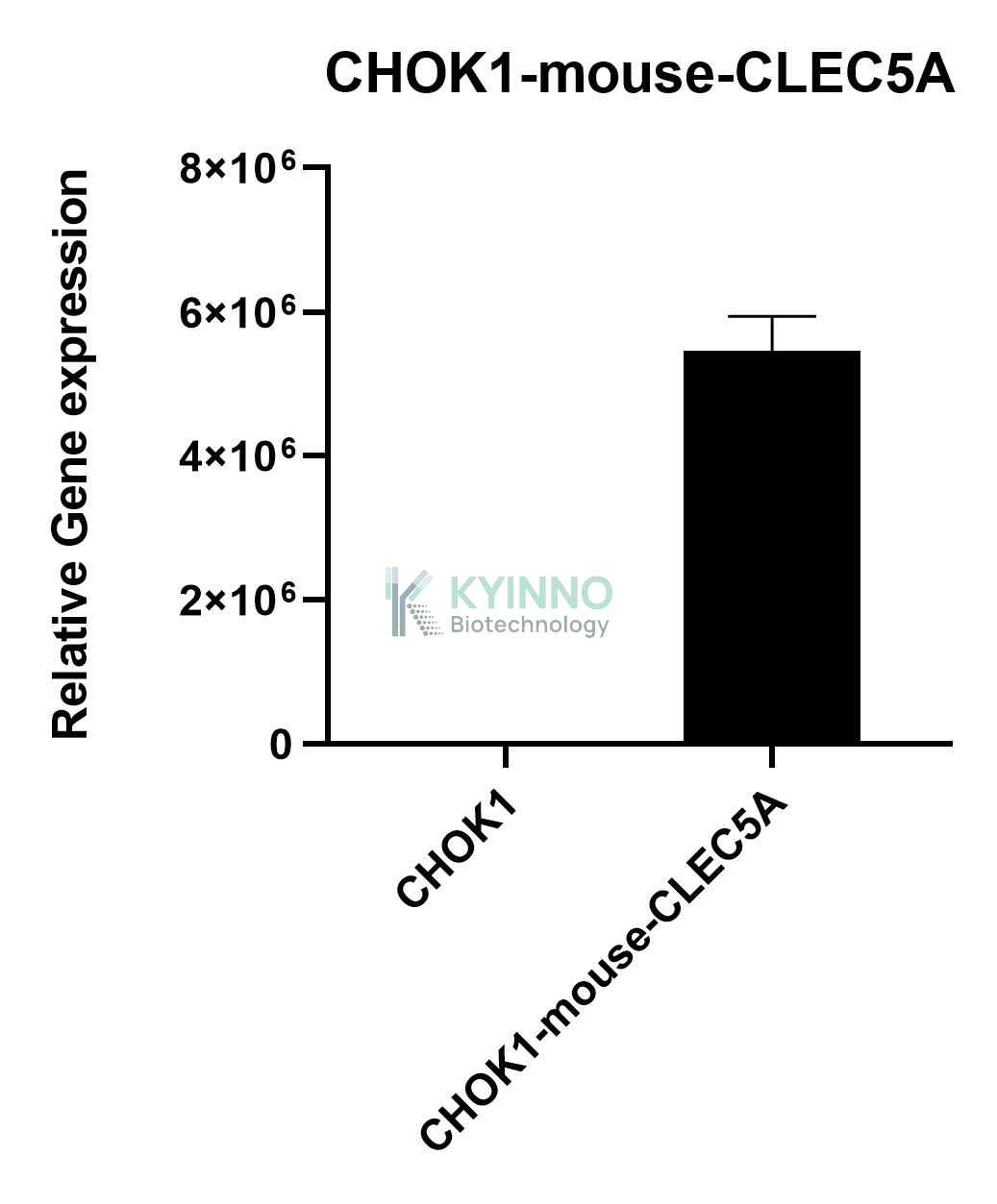
Figure 1: Characterization of mouse CLEC5A overexpression in the CHOK1 mouse CLEC5A stable clone using qPCR.

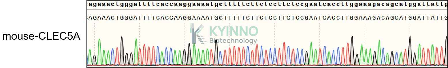
Figure 2: Characterization of mouse CLEC5A overexpression in CHOK1 stable clones using DNA sequencing.

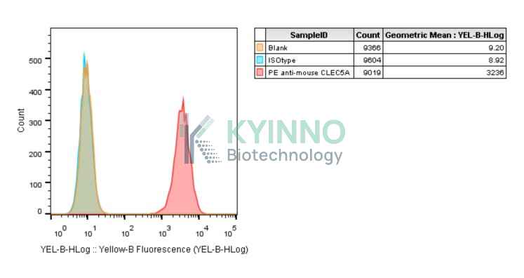
Hybridoma or ligand binding screening with FACS.

Cell Resuscitation

  1. Prewarm culture medium (RPMI1640 supplemented with 10% FBS and 10μg/mL Puromycin)in a 37°C water bath.
  2. Thaw the frozen vial in a 37°C water bath for 1-2 minutes.
  3. Transfer the vial into biosafety cabinet, and wipe the surface with 70% ethanol.
  4. Unscrew the top of the vial and transfer the cell suspension gently into a sterile centrifuge tube containing 9.0mL complete culture medium.
  5. Spin at ~ 125 × g for 5-7 minutes at room temperature, and discard the supernatant without disturbing the pellet.
  6. Resuspend cell pellet with the appropriate volume of complete medium and transfer the cell suspension into a T25 culture flask.
  7. Incubate the flask at 37°C, 5% CO2 incubator.
  8. Split saturated culture 1:4-1:8 every 2-3 days; seed out at about 1-3 × 10⁵ cells/mL.

Cell Freezing

  1. Prepare the freezing medium (70% RPMI1640 + 20% FBS + 10% DMSO) fresh immediately before use.
  2. Keep the freezing medium on ice and label cryovials.
  3. Transfer cells to a sterile, conical centrifuge tube, and count the cells.
  4. Centrifuge the cells at 250×g for 5 minutes at room temperature and carefully aspirate off the medium.
  5. Resuspend the cells at a density of at least 3×10⁶ cells/mL in chilled freezing medium.
  6. Aliquot 1 mL of the cell suspension into each cryovial.
  7. Freeze cells in the CoolCell freezing container overnight in a -80°C freezer.
  8. Transfer vials to liquid nitrogen for long-term storage

References

  1. Aoki N, Kimura Y, Kimura S, Nagato T, Azumi M, Kobayashi H, Sato K, Tateno M. Expression and functional role of MDL-1 (CLEC5A) in mouse myeloid lineage cells. J Leukoc Biol. 2009 Mar;85(3):508-17. doi: 10.1189/jlb.0508329. Epub 2008 Dec 12. PMID: 19074552.
Please enable JavaScript in your browser to complete this form.