KC-3911

Daudi-Luc2-EGFP Cell Line

×
Please enable JavaScript in your browser to complete this form.
35018
Home » 细胞系 » Daudi-Luc2-EGFP Cell Line

Background of Daudi-Luc2-EGFP Cell Line

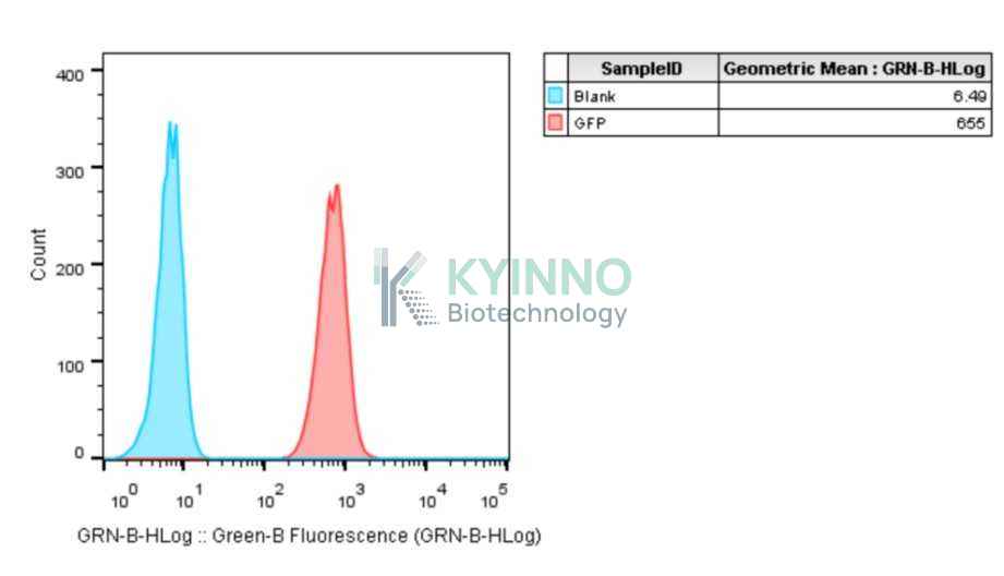
Green fluorescence protein(GFP) is a protein composed with 238 amino acids and isolated from Aequorea victoria. GFP emits green fluorescence spontaneously. 
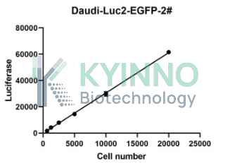
Luciferase is an oxidative enzyme that can produce bioluminescence with the addition of luciferin, but don’t need an external light source, which is different from fluorescent proteins. The bioluminescence can be detected directly by light sensitive device, such as luminometer or modified microscope. Luciferase is widely used in many fields of biological research, such as transcriptional activity, kinase or other enzyme activity, cellular ATP level, whole animal imaging.

Specifications

Catalog NumberKC-3911
Cell Line NameDaudi-Luc2-EGFP Cell Line
Host Cell LineDaudi
DescriptionStable Daudi cell line expressing exogenous luciferase and eGFP gene
QuantityTwo vials of frozen cells (≥2-106/vial)
StabilityStable in culture over a minimum of 10 passages
ApplicationDrug screening and biological assays
Freezing Medium70% RPMI1640 + 20% FBS + 10% DMSO
Propagation MediumRPMI1640+10%FBS+600µg/mL Hygromycin B
Selection MarkerHygromycin B
Morphologylymphoblastic cell cells growing as a Suspended
SubcultureSplit saturated culture 1:4-1:5 every 2-3 days; seed out at about 1-3 × 10⁵ cells/mL
Incubation37 °C with 5% CO2
StorageLiquid nitrogen immediately upon receiving
Doubling TimeApproximately 30 hours
Mycoplasma StatusNegative
In Vivo ValidationNA

Cell Line Generation

Daudi Luc2 EGFP cell line was generated using a lentiviral vector expressing the luciferase and EGFP sequence.

Characterization

Figure 1: Characterization of EGFP overexpression in the Daudi Luc2 EGFP stable clone using FACS.

Figure 2: Characterization of luciferase expression in Daudi Luc2 EGFP stable clone using the Bright-Lite Luciferase Detection System.

Cell Resuscitation

1. Prewarm culture medium (RPMI1640 supplemented with 10% FBS and 600µg/mL Hygromycin B)in a 37°C water bath. 2. Thaw the frozen vial in a 37°C water bath for 1-2 minutes. 3. Transfer the vial into biosafety cabinet, and wipe the surface with 70% ethanol. 4. Unscrew the top of the vial and transfer the cell suspension gently into a sterile centrifuge tube containing 9.0mL complete culture medium. 5. Spin at ~ 125 × g for 5-7 minutes at room temperature, and discard the supernatant without disturbing the pellet. 6. Resuspend cell pellet with the appropriate volume of complete medium and transfer the cell suspension into a T25 culture flask. 7. Incubate the flask at 37°C, 5% CO2 incubator. 8. Split saturated culture 1:4-1:5 every 2-3 days; seed out at about 1-3 × 10⁵ cells/mL.

Cell Freezing

1. Prepare the freezing medium (70% RPMI1640 + 20% FBS + 10% DMSO) fresh immediately before use. 2. Keep the freezing medium on ice and label cryovials. 3. Harvest cells in a sterile, conical centrifuge tube during the logarithmic growth period and count the cells. 4. Centrifuge the cells at 250×g for 5 minutes at room temperature and carefully aspirate off the medium. 5. Resuspend the cells at a density of at least 3×10⁶ cells/mL in chilled freezing medium. 6. Aliquot 1 mL of the cell suspension into each cryovial. 7. Freeze cells in the CoolCell freezing container overnight in a -80°C freezer. 8. Transfer vials to liquid nitrogen for long-term storage.

References

1. Greer LF 3rd, Szalay AA. Imaging of light emission from the expression of luciferases in living cells and organisms: a review. Luminescence. 2002 Jan-Feb; 17(1): 43-74. doi: 10.1002/bio. 676. PMID: 11816060. 2. Falkowska-Hansen B, Kollar J, Grüner BM, et al. An inducible Tet-Off-H2B-GFP lentiviral reporter vector for detection and in vivo isolation of label-retaining cells. Exp Cell Res. 2010 Jul 1; 316(11): 1885-95. doi: 10.1016/j. yexcr. 2010.02.015. Epub 2010 Feb 18. PMID: 20171964.
Please enable JavaScript in your browser to complete this form.