KC-3984

Jurkat-NFAT-Luc2-CD7-KO-3A4 Cell Line

×
Please enable JavaScript in your browser to complete this form.
40426
Home » 细胞系 » Jurkat-NFAT-Luc2-CD7-KO-3A4 Cell Line

Background of Jurkat-NFAT-Luc2-CD7-KO-3A4 Cell Line

CD7 is located on chromosome 17 and encodes a cell surface glycoprotein member of the immunoglobulin superfamily. The protein is found on thymocytes and mature NK and T-cells. It plays an essential role in T-cell interactions and also in T-cell/B-cell interactions during early lymphopoeisis. CD7 is highly expressed on the surface of T-ALL/LBL T cells and is considered a viable CAR-T therapeutic target. Although CD7 is expressed on natural killer (NK) cells and normal T cells at various stages of maturation, CD7 knockout mice develop normal lymphoid organs and immune responses, suggesting that CD7 is dispensable for T/NK cell development and functions.

Specifications

Catalog NumberKC-3984
Cell Line NameJurkat-NFAT-Luc2-CD7-KO-3A4 Cell Line
Host Cell LineJurkat-NFAT-Luc2
DescriptionStable Jurkat-NFAT-Luc2-reporter cell line with CD7 gene knockout
QuantityTwo vials of frozen cells (≥2-106/vial)
StabilityStable in culture over a minimum of 10 passages
ApplicationDrug screening and biological assays
Freezing MediumRPMI1640+20% FBS+10% DMSO
Propagation MediumRPMI1640+10% FBS+300μg/ml Hygromycin B
Selection MarkerNA
MorphologyLymphoblast
SubcultureSplit saturated culture 1:4-1:5 every 2-3 days; seed out at about 1-3 × 10⁵ cells/mL
Incubation37 °C with 5% CO2
StorageLiquid nitrogen immediately upon receiving
Doubling TimeApproximately 30 hours
Mycoplasma StatusNegative
In Vivo ValidationNA

Cell Line Generation

Jurkat-NFAT-Luc2-CD7-KO-3A4 cell line was generated using the CRISPR method.

Characterization

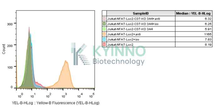
Figure 1: Characterization of Jurkat-NFAT-Luc2-CD7-KO Cell Line stable clone using FACS.

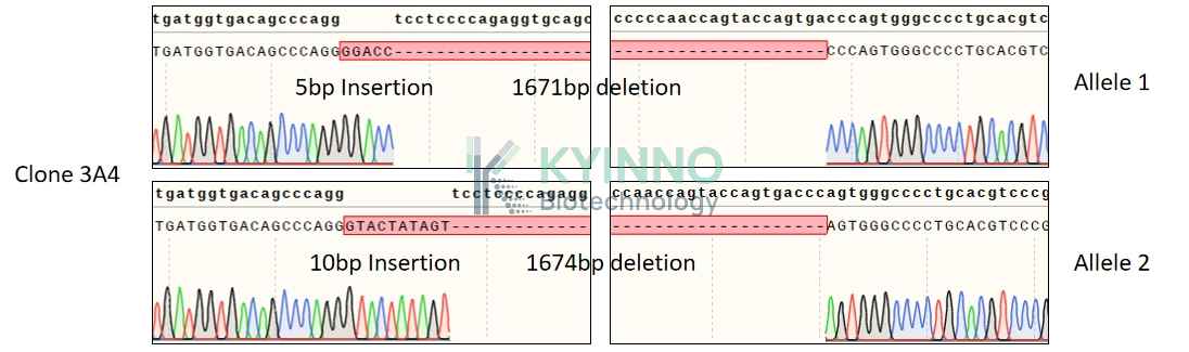
Figure 2: Characterization of Jurkat-NFAT-Luc2-CD7-KO-3A4 cell line stable clone using PCR sequencing.

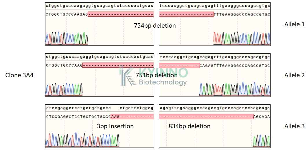
Figure 3: Characterization of Jurkat-NFAT-Luc2-CD7-KO-3A4 cell line stable clone using RT-PCR sequencing.

Cell Resuscitation

  1. Prewarm culture medium (RPMI1640+10% FBS+300μg/ml Hygromycin B)in a 37°C water bath.
  2. Thaw the frozen vial in a 37°C water bath for 1-2 minutes.
  3. Transfer the vial into biosafety cabinet, and wipe the surface with 70% ethanol.
  4. Unscrew the top of the vial and transfer the cell suspension gently into a sterile centrifuge tube containing 9.0mL complete culture medium.
  5. Spin at ~ 125 × g for 5-7 minutes at room temperature, and discard the supernatant without disturbing the pellet.
  6. Resuspend cell pellet with the appropriate volume of complete medium and transfer the cell suspension into a T25 culture flask.
  7. Incubate the flask at 37°C, 5% CO2 incubator.
  8. Split saturated culture 1:4-1:5 every 2-3 days; seed out at about 1-3 × 10⁵ cells/mL.

Cell Freezing

  1. Prepare the freezing medium (70% RPMI-1640 + 20% FBS + 10% DMSO) fresh immediately before use.
  2. Keep the freezing medium on ice and label cryovials.
  3. Transfer cells to a sterile, conical centrifuge tube, and count the cells.
  4. Centrifuge the cells at 250×g for 5 minutes at room temperature and carefully aspirate off the medium.
  5. Resuspend the cells at a density of at least 3×10⁶ cells/mL in chilled freezing medium.
  6. Aliquot 1 mL of the cell suspension into each cryovial.
  7. Freeze cells in the CoolCell freezing container overnight in a -80°C freezer.
  8. Transfer vials to liquid nitrogen for long-term storage.

References

  1. Rogers SL, Zhao Y, Jiang X, Eaves CJ, Mager DL, Rouhi A. Expression of the leukemic prognostic marker CD7 is linked to epigenetic modifications in chronic myeloid leukemia. Mol Cancer. 2010 Feb 22;9:41. doi: 10.1186/1476-4598-9-41. PMID: 20175919; PMCID: PMC2843654.
  2. Lu P, Liu Y, Yang J, Zhang X, Yang X, Wang H, Wang L, Wang Q, Jin D, Li J, Huang X. Naturally selected CD7 CAR-T therapy without genetic manipulations for T-ALL/LBL: first-in-human phase 1 clinical trial. Blood. 2022 Jul 28;140(4):321-334. doi: 10.1182/blood.2021014498. PMID: 35500125.
Please enable JavaScript in your browser to complete this form.