KC-4173

Jurkat-CD3D-CD3E-KO-2A2-Cell-Line

×
请在浏览器中启用JavaScript来完成此表单。
42387
Home » Jurkat-CD3D-CD3E-KO-2A2-Cell-Line

Background of Jurkat-CD3D-CD3E-KO-2A2-Cell-Line

The protein encoded by CD3D is part of the T-cell receptor/CD3 complex (TCR/CD3 complex) and is involved in T-cell development and signal transduction. The encoded membrane protein represents the delta subunit of the CD3 complex, and along with four other CD3 subunits, binds either TCR alpha/beta or TCR gamma/delta to form the TCR/CD3 complex on the surface of T-cells. Defects in this gene are a cause of severe combined immunodeficiency autosomal recessive T-cell-negative/B-cell-positive/NK-cell-positive (SCIDBNK).
The protein encoded by CD3E is the CD3-epsilon polypeptide, which together with CD3-gamma, -delta and -zeta, and the T-cell receptor alpha/beta and gamma/delta heterodimers, forms the T-cell receptor-CD3 complex. This complex plays an important role in coupling antigen recognition to several intracellular signal-transduction pathways. The genes encoding the epsilon, gamma and delta polypeptides are located in the same cluster on chromosome 11. The epsilon polypeptide plays an essential role in T-cell development. Defects in this gene cause immunodeficiency.

Specifications

Catalog NumberKC-4173
Cell Line NameJurkat-CD3D-CD3E-KO-2A2-Cell-Line
Host Cell LineJurkat-CD3E-KO-1A1
DescriptionStable Jurkat-CD3E-KO clone with human CD3D gene knockout, No.2A2
QuantityOne vial of frozen cells (≥2-106/vial)
StabilityStable in culture over a minimum of 10 passages
ApplicationDrug screening and biological assays
Freezing MediumRPMI1640+20% FBS+10% DMSO
Propagation MediumRPMI1640+10% FBS
Selection MarkerN/A
MorphologyLymphoblast
SubcultureSplit saturated culture 1:4-1:6 every 2-3 days; seed out at about 1-3 × 10⁵ cells/mL
Incubation37 °C with 5% CO2
StorageLiquid nitrogen immediately upon receiving
Doubling TimeApproximately 26 hours
Mycoplasma StatusNegative
In Vivo ValidationNA

Cell Line Generation

Characterization

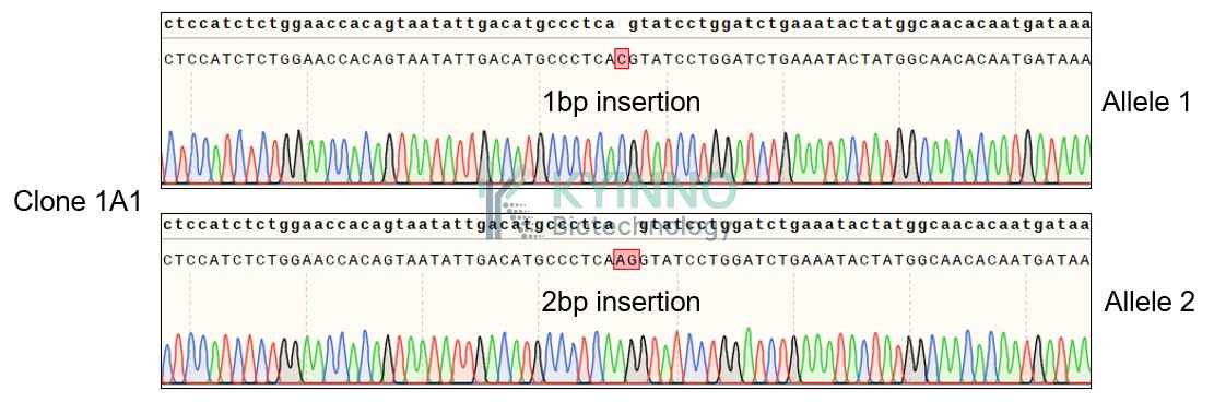
Figure 1: Characterization of CD3E knockout in Jurkat using PCR sequencing.

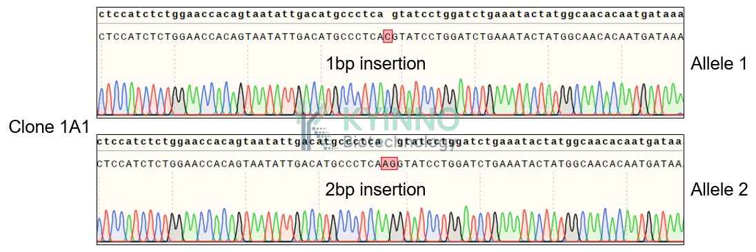
Figure 2: Characterization of CD3E knockout in Jurkat using RT-PCR sequencing.

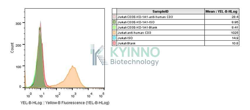
Figure 3: Characterization of CD3E knockout in Jurkat using FACS.

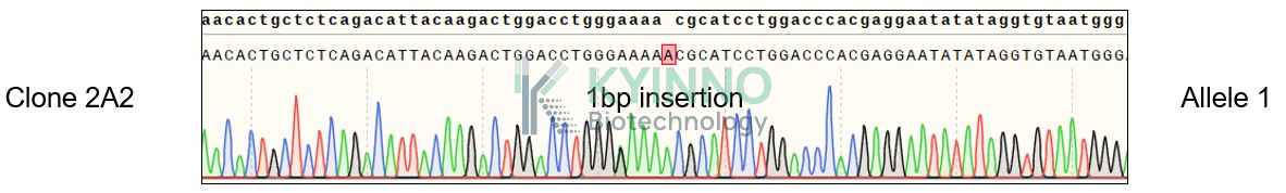
Figure 4: Characterization of CD3D knockout in Jurkat-CD3E-KO using PCR sequencing.

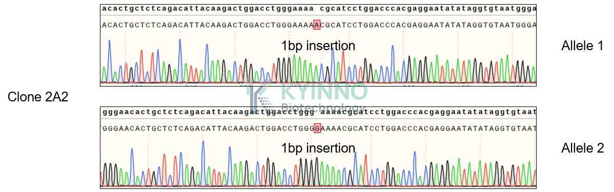
Figure 5: Characterization of CD3D knockout in Jurkat-CD3E-KO using RT-PCR sequencing.

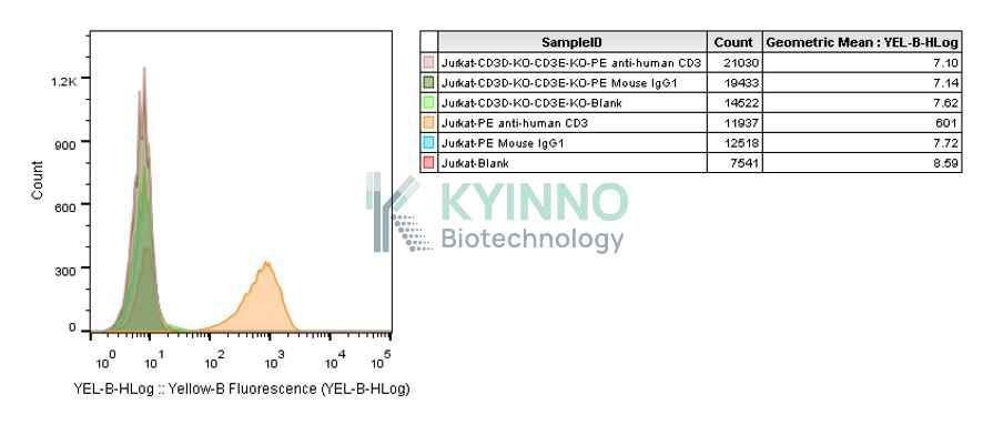
Figure 6: Characterization of CD3D knockout in Jurkat-CD3E-KO usingFACS.

Cell Resuscitation

Cell Freezing

References

1.https://www.ncbi.nlm.nih.gov/gene/915
2.https://www.ncbi.nlm.nih.gov/gene/916

Use License Agreement

Research Use Only.
Not for use in diagnostic procedures or therapeutic applications.
Redistribution of the cell line or its derivatives is prohibited without prior written permission from Kyinno Biotechnology.
请在浏览器中启用JavaScript来完成此表单。