KC-4409

T47D-RB1-KO-1A1-Cell-Line

×
Please enable JavaScript in your browser to complete this form.
46096
Home » T47D-RB1-KO-1A1-Cell-Line

Background of T47D-RB1-KO-1A1-Cell-Line

The Retinoblastoma 1 (RB1) gene, located on chromosome 13q14, is a tumor suppressor gene essential for cell cycle regulation. The protein encoded by this gene is a negative regulator of the cell cycle and was the first tumor suppressor gene found. The encoded protein also stabilizes constitutive heterochromatin to maintain the overall chromatin structure. The active, hypophosphorylated form of the protein binds transcription factor E2F1. Defects in this gene are a cause of childhood cancer retinoblastoma (RB), bladder cancer, and osteogenic sarcoma.

Specifications

Catalog NumberKC-4409
Cell Line NameT47D-RB1-KO-1A1-Cell-Line
Host Cell LineT47D
DescriptionStable T47D clone with human RB1 gene knockout, No.1A1
QuantityTwo vials of frozen cells (≥2-106/vial)
StabilityStable in culture over a minimum of 10 passages
ApplicationDrug screening and biological assays
Freezing MediumRPMI1640+20% FBS+10% DMSO
Propagation MediumRPMI1640+10% FBS+0.2 Units/mL Insulin
Selection MarkerN/A
Morphologyepithelial
SubcultureSplit saturated culture 1:3-1:5 every 2-3 days; seed out at about 1-3 × 10⁵ cells/mL
Incubation37 °C with 5% CO2
StorageLiquid nitrogen immediately upon receiving
Doubling TimeApproximately 60 hours
Mycoplasma StatusNegative
In Vivo ValidationNA

Cell Line Generation

T47D-RB1-KO-1A1 cell line was generated using the CRISPR method.

Characterization

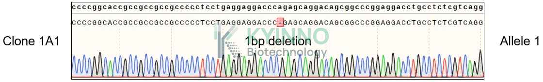
Figure 1: Characterization of T47D-RB1-KO-1A1 cell line stable clone using PCR sequencing.

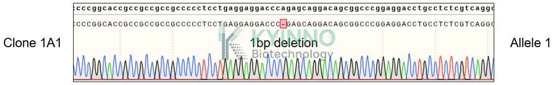
Figure 2: Characterization of T47D-RB1-KO-1A1 cell line stable clone using RT-PCR sequencing.

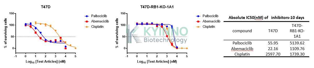
Figure 3: Characterization of Dose-response curves and IC50 values for T47D and T47D-RB1-KO-1A1 cells treated with Palbociclib, Abemaciclib, and Cisplatin over 5 days.

Cell Resuscitation

  1. Prewarm culture medium (RPMI1640 + 10% FBS+0.2 Units/mL Insulin)in a 37°C water bath.
  2. Thaw the frozen vial in a 37°C water bath for 1-2 minutes.
  3. Transfer the vial into biosafety cabinet, and wipe the surface with 70% ethanol.
  4. Unscrew the top of the vial and transfer the cell suspension gently into a sterile centrifuge tube containing 9.0mL complete culture medium.
  5. Spin at ~ 125 × g for 5-7 minutes at room temperature, and discard the supernatant without disturbing the pellet.
  6. Resuspend cell pellet with the appropriate volume of complete medium and transfer the cell suspension into a T25 culture flask.
  7. Incubate the flask at 37°C, 5% CO2 incubator.
  8. Split saturated culture 1:3-1:5 every 2-3 days; seed out at about 1-3 × 10⁵ cells/mL.

Cell Freezing

  1. Prepare the freezing medium (70% RPMI-1640 + 20% FBS + 10% DMSO) fresh immediately before use.
  2. Keep the freezing medium on ice and label cryovials.
  3. Transfer cells to a sterile, conical centrifuge tube, and count the cells.
  4. Centrifuge the cells at 250×g for 5 minutes at room temperature and carefully aspirate off the medium.
  5. Resuspend the cells at a density of at least 3×10⁶ cells/mL in chilled freezing medium.
  6. Aliquot 1 mL of the cell suspension into each cryovial.
  7. Freeze cells in the CoolCell freezing container overnight in a -80°C freezer.
  8. Transfer vials to liquid nitrogen for long-term storage

References

  1. Classon M, Harlow E. The retinoblastoma tumour suppressor in development and cancer. Nat Rev Cancer. 2002 Dec;2(12):910-7. doi: 10.1038/nrc950. PMID: 12459729. 2.Knudsen ES, Wang JY. Targeting the RB-pathway in cancer therapy. Clin Cancer Res. 2010 Feb 15;16(4):1094-9. doi: 10.1158/1078-0432.CCR-09-0787. Epub 2010 Feb 9. PMID: 20145169; PMCID: PMC2822892. 3.https://www.ncbi.nlm.nih.gov/gene/5925
Please enable JavaScript in your browser to complete this form.