KC-4425

HCT116-WRN-C727S-KI-3A1 Cell Line

×
Please enable JavaScript in your browser to complete this form.
47786
Home » HCT116-WRN-C727S-KI-3A1 Cell Line

Background of HCT116-WRN-C727S-KI-3A1 Cell Line

The WRN gene, encoding the Werner syndrome RecQ-like helicase (WRN), is a member of the RecQ helicase family. This protein plays a crucial role in maintaining genomic stability through its involvement in DNA repair, replication, and telomere maintenance. Mutations in the WRN gene are responsible for Werner syndrome, a rare autosomal recessive disorder characterized by premature aging and an increased risk of cancer. Individuals with Werner syndrome exhibit symptoms such as early onset of atherosclerosis, type 2 diabetes, osteoporosis, and cataracts, typically developing these conditions in their 20s or 30s.

Specifications

Catalog NumberKC-4425
Cell Line NameHCT116-WRN-C727S-KI-3A1 Cell Line
Host Cell LineHCT116
DescriptionStable HCT116 clone expressing exogenous WRN gene bearing C727S mutations, No.3A1
QuantityTwo vials of frozen cells (≥2-106/vial)
StabilityStable in culture over a minimum of 10 passages
ApplicationDrug screening and biological assays
Freezing MediumMcCoy‘s 5a+20% FBS+10% DMSO
Propagation MediumMcCoy‘s 5a+10% FBS
Selection MarkerN/A
MorphologyEpithelial
SubcultureSplit saturated culture 1:3-1:6 every 2-3 days; seed out at about 1-3 × 10⁵ cells/mL
Incubation37 °C with 5% CO2
StorageLiquid nitrogen immediately upon receiving
Doubling TimeApproximately 30 hours
Mycoplasma StatusNegative

Cell Line Generation

HCT116-WRN-C727S-3A1 cell line was generated using the CRISPR method.

Characterization

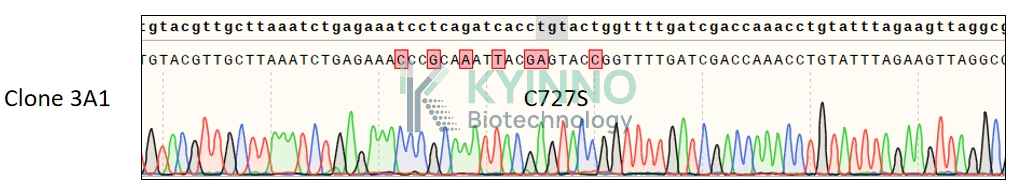
Figure 1: Characterization of HCT116-WRN-C727S-KI-3A1 cell line stable clone using PCR sequencing.

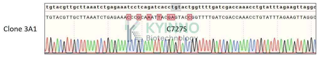
Figure 2: Characterization of HCT116-WRN-C727S-KI-3A1 cell line stable clone using RT-PCR sequencing.

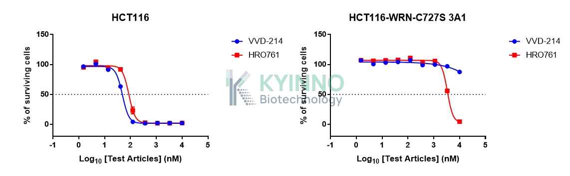
Figure 3. Characterization of dose-response curves for WRN inhibitors on HCT116 and HCT116-WRN-C727S-KI-3A1 cells.

Cell Resuscitation

  1. Prewarm culture medium (McCoy‘s 5a + 10% FBS)in a 37°C water bath.
  2. Thaw the frozen vial in a 37°C water bath for 1-2 minutes.
  3. Transfer the vial into biosafety cabinet, and wipe the surface with 70% ethanol.
  4. Unscrew the top of the vial and transfer the cell suspension gently into a sterile centrifuge tube containing 9.0mL complete culture medium.
  5. Spin at ~ 125 × g for 5-7 minutes at room temperature, and discard the supernatant without disturbing the pellet.
  6. Resuspend cell pellet with the appropriate volume of complete medium and transfer the cell suspension into a T25 culture flask.
  7. Incubate the flask at 37°C, 5% CO2 incubator.
  8. Split saturated culture 1:3-1:6 every 2-3 days; seed out at about 1-3 × 10⁵ cells/mL.

Cell Freezing

  1. Prepare the freezing medium (70% McCoy‘s 5a + 20% FBS + 10% DMSO) fresh immediately before use.
  2. Keep the freezing medium on ice and label cryovials.
  3. Transfer cells to a sterile, conical centrifuge tube, and count the cells.
  4. Centrifuge the cells at 250×g for 5 minutes at room temperature and carefully aspirate off the medium.
  5. Resuspend the cells at a density of at least 3×10⁶ cells/mL in chilled freezing medium.
  6. Aliquot 1 mL of the cell suspension into each cryovial.
  7. Freeze cells in the CoolCell freezing container overnight in a -80°C freezer.
  8. Transfer vials to liquid nitrogen for long-term storage.

References

  1. Opresko, P. L., et al. (2014). The Werner syndrome helicase and exonuclease cooperate to resolve telomeric D loops in a manner regulated by TRF1 and TRF2.Molecular Cell, 54(6), 1163-1174. https://doi.org/10.1016/j.molcel.2014.05.018
  2. Monnat, R. J., Jr. (2010). Human RECQ helicases: roles and mechanisms.Nature Reviews Molecular Cell Biology, 11(10), 721-733. https://doi.org/10.1038/nrm2970
  3. Oshima, J., et al. (2017). Werner syndrome: a review on clinical features, molecular mechanisms, and potential therapies.Aging and Disease, 8(5), 624-644. https://doi.org/10.14336/AD.2017.0115
Please enable JavaScript in your browser to complete this form.