KC-4711

293T STAT1 Luc2 Cell Line

×
Please enable JavaScript in your browser to complete this form.
49075
Home » 293T STAT1 Luc2 Cell Line

Background of 293T STAT1 Luc2 Cell Line

Signal transducer and transcription activator that mediates cellular responses to interferons (IFNs), cytokine KITLG/SCF and other cytokines and other growth factors. Following type I IFN (IFN-alpha and IFN-beta) binding to cell surface receptors, signaling via protein kinases leads to activation of Jak kinases (TYK2 and JAK1) and to tyrosine phosphorylation of STAT1 and STAT2. The phosphorylated STATs dimerize and associate with ISGF3G/IRF-9 to form a complex termed ISGF3 transcription factor, that enters the nucleus. ISGF3 binds to the IFN stimulated response element (ISRE) to activate the transcription of IFN-stimulated genes (ISG), which drive the cell in an antiviral state. In response to type II IFN (IFN-gamma), STAT1 is tyrosine- and serine-phosphorylated. It then forms a homodimer termed IFN-gamma-activated factor (GAF), migrates into the nucleus and binds to the IFN gamma activated sequence (GAS) to drive the expression of the target genes, inducing a cellular antiviral state. Becomes activated in response to KITLG/SCF and KIT signaling. May mediate cellular responses to activated FGFR1, FGFR2, FGFR3 and FGFR4. Following bacterial lipopolysaccharide (LPS)-induced TLR4 endocytosis, phosphorylated at Thr-749 by IKBKB which promotes binding of STAT1 to the 5'-TTTGAGGC-3' sequence in the ARID5A promoter, resulting in transcriptional activation of ARID5A and subsequent ARID5A-mediated stabilization of IL6. Phosphorylation at Thr-749 also promotes binding of STAT1 to the 5'-TTTGAGTC-3' sequence in the IL12B promoter and activation of IL12B transcription. Involved in food tolerance in small intestine: associates with the Gasdermin-D, p13 cleavage product (13 kDa GSDMD) and promotes transcription of CIITA, inducing type 1 regulatory T (Tr1) cells in upper small intestine (By similarity).

Specifications

Catalog NumberKC-4711
Cell Line Name293T STAT1 Luc2 Cell Line
Host Cell Line293T
DescriptionStable 293T cell line expressing exogenous luciferase gene under the control of STAT1 signaling pathway.
QuantityTwo vials of frozen cells (≥2-106/vial)
StabilityStable in culture over a minimum of 10 passages
ApplicationDrug screening and biological assays
Freezing Medium70% DMEM+20% FBS+10% DMSO
Propagation MediumDMEM+10% FBS +150μg/mL Hygromycin B + 1μg/mL Puromycin
Selection MarkerHygromycin B and Puromycin
MorphologyEpithelial
SubcultureSplit saturated culture 1:4-1:5 every 2-3 days; seed out at about 1-3 × 10⁵ cells/mL
Incubation37 °C with 5% CO2
StorageLiquid nitrogen immediately upon receiving
Doubling TimeApproximately 30 hours
Mycoplasma StatusNegative

Cell Line Generation

293T-STAT1-Luc2 cell line was generated using lentivirus expressing STAT1-Luc2 and STAT1 sequence.

Characterization

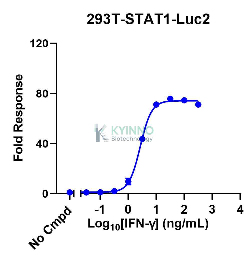
Figure: 293T-STAT1-Luc2 cells were seeded into the 96-well plate, treated with IFN-γ for 16 hours, then readout with Bright-lite™ Luciferase Assay system.

Cell Resuscitation

  1. Prewarm culture medium (DMEM supplemented with 10% FBS, 150μg/mL Hygromycin B and 1μg/mL Puromycin)in a 37°C water bath.
  2. Thaw the frozen vial in a 37°C water bath for 1-2 minutes.
  3. Transfer the vial into biosafety cabinet, and wipe the surface with 70% ethanol.
  4. Unscrew the top of the vial and transfer the cell suspension gently into a sterile centrifuge tube containing 9.0mL complete culture medium.
  5. Spin at ~ 125 × g for 5-7 minutes at room temperature, and discard the supernatant without disturbing the pellet.
  6. Resuspend cell pellet with the appropriate volume of complete medium and transfer the cell suspension into a T25 culture flask.
  7. Incubate the flask at 37°C, 5% CO2 incubator.
  8. Split saturated culture 1:4-1:8 every 2-3 days; seed out at about 1-3 × 10⁵ cells/mL.

Cell Freezing

  1. Prepare the freezing medium (70% DMEM + 20% FBS + 10% DMSO) fresh immediately before use.
  2. Keep the freezing medium on ice and label cryovials.
  3. Transfer cells to a sterile, conical centrifuge tube, and count the cells.
  4. Centrifuge the cells at 250×g for 5 minutes at room temperature and carefully aspirate off the medium.
  5. Resuspend the cells at a density of at least 3×10⁶ cells/mL in chilled freezing medium.
  6. Aliquot 1 mL of the cell suspension into each cryovial.
  7. Freeze cells in the CoolCell freezing container overnight in a -80°C freezer.
  8. Transfer vials to liquid nitrogen for long-term storage

References

1. Rogers RS, Horvath CM, Matunis MJ. SUMO modification of STAT1 and its role in PIAS-mediated inhibition of gene activation. J Biol Chem. 2003 Aug 8;278(32):30091-7. doi: 10.1074/jbc.M301344200. Epub 2003 May 22. PMID: 12764129.
2. Zhang Y, Mao D, Roswit WT, Jin X, Patel AC, Patel DA, Agapov E, Wang Z, Tidwell RM, Atkinson JJ, Huang G, McCarthy R, Yu J, Yun NE, Paessler S, Lawson TG, Omattage NS, Brett TJ, Holtzman MJ. PARP9-DTX3L ubiquitin ligase targets host histone H2BJ and viral 3C protease to enhance interferon signaling and control viral infection. Nat Immunol. 2015 Dec;16(12):1215-27. doi: 10.1038/ni.3279. Epub 2015 Oct 19. PMID: 26479788; PMCID: PMC4653074.
Please enable JavaScript in your browser to complete this form.