KC-4639

Jurkat-NFAT-Luc2-ACVR2B-KO-CD16-V158-OE Cell Line

×
Please enable JavaScript in your browser to complete this form.
52156
Home » Jurkat-NFAT-Luc2-ACVR2B-KO-CD16-V158-OE Cell Line

Background of Jurkat-NFAT-Luc2-ACVR2B-KO-CD16-V158-OE Cell Line

ACVR2B, also known as Activin A Receptor Type IIB or ActR-IIB, is a transmembrane serine/threonine kinase receptor encoded by the ACVR2B gene. It belongs to the Transforming Growth Factor-β (TGF-β) superfamily and plays a crucial role in various biological processes. ACVR2B is expressed in multiple tissues, with high levels in tumor cells. It binds to Activin A, recruiting and phosphorylating type I activin receptors, and subsequently signals through SMAD2/3 proteins to regulate cell proliferation, differentiation, and migration. Mutations in ACVR2B have been associated with left-right axis malformations and heterotaxy syndrome.

Specifications

Catalog NumberKC-4639
Cell Line NameJurkat-NFAT-Luc2-ACVR2B-KO-CD16-V158-OE Cell Line
Host Cell LineJurkat-NFAT-Luc2-CD16-V158-OE
DescriptionStable Jurkat-NFAT-Luc2-CD16-V158-OE clone with human ACVR2B gene knockout, No.1A3
QuantityTwo vials of frozen cells (≥2-106/vial)
StabilityStable in culture over a minimum of 10 passages
ApplicationDrug screening and biological assays
Freezing MediumRPMI1640+20% FBS+10% DMSO
Propagation MediumRPMI1640+10% FBS+0.75μg/mL Puromycin+300μg/mL Hygromycin B
Selection MarkerPuromycin, Hygromycin B
MorphologyLymphoblast
SubcultureSplit saturated culture 1:4-1:6 every 2-3 days; seed out at about 1-3 × 10⁵ cells/Ml
Incubation37 °C with 5% CO2
StorageLiquid nitrogen immediately upon receiving
Doubling TimeApproximately 26 hours
Mycoplasma StatusNegative

Cell Line Generation

Jurkat-NFAT-Luc2-ACVR2B-KO-CD16-V158-OE cell line was generated using the CRISPR method.

Characterization

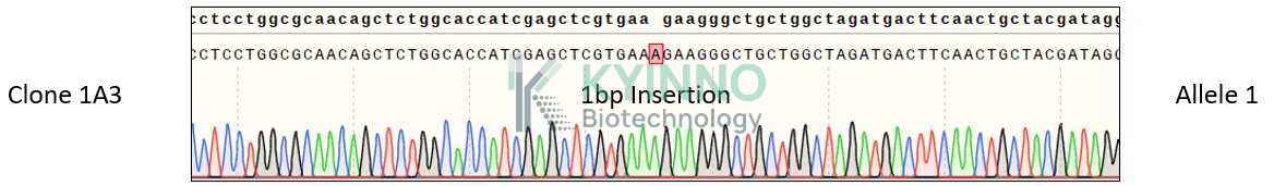
Figure 1: Characterization of ACVR2B knockout in Jurkat-NFAT-Luc2-CD16-V158-OE using PCR sequencing.

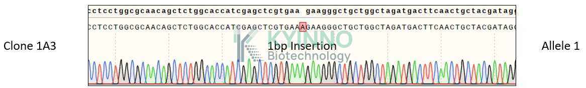
Figure 2: Characterization of ACVR2B knockout in Jurkat-NFAT-Luc2-CD16-V158-OE using RT-PCR sequencing.

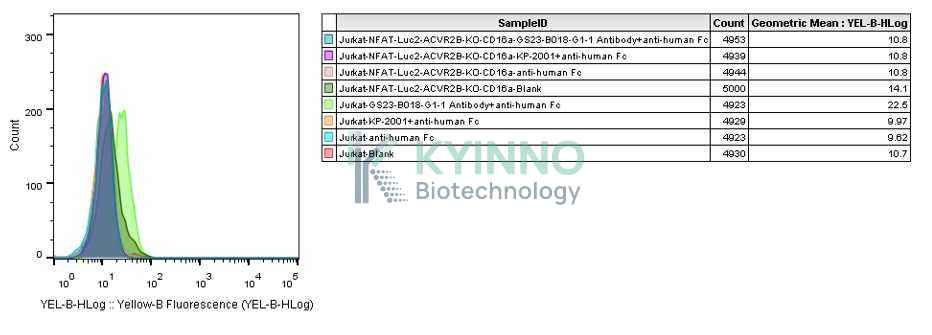
Figure 3: Characterization of ACVR2B knockout in Jurkat-NFAT-Luc2-CD16-V158-OE using FACS.

Cell Resuscitation

  1. Prewarm culture medium (RPMI1640+10% FBS+0.75μg/mL Puromycin+300μg/mL Hygromycin B)in a 37°C water bath.
  2. Thaw the frozen vial in a 37°C water bath for 1-2 minutes.
  3. Transfer the vial into biosafety cabinet, and wipe the surface with 70% ethanol.
  4. Unscrew the top of the vial and transfer the cell suspension gently into a sterile centrifuge tube containing 9.0mL complete culture medium.
  5. Spin at ~ 125 × g for 5-7 minutes at room temperature, and discard the supernatant without disturbing the pellet.
  6. Resuspend cell pellet with the appropriate volume of complete medium and transfer the cell suspension into a T25 culture flask.
  7. Incubate the flask at 37°C, 5% CO2 incubator.
  8. Split saturated culture 1:4-1:6 every 2-3 days; seed out at about 1-3 × 10⁵ cells/mL.

Cell Freezing

  1. Prepare the freezing medium (70% RPMI-1640 + 20% FBS + 10% DMSO) fresh immediately before use.
  2. Keep the freezing medium on ice and label cryovials.
  3. Transfer cells to a sterile, conical centrifuge tube, and count the cells.
  4. Centrifuge the cells at 250×g for 5 minutes at room temperature and carefully aspirate off the medium.
  5. Resuspend the cells at a density of at least 3×10⁶ cells/mL in chilled freezing medium.
  6. Aliquot 1 mL of the cell suspension into each cryovial.
  7. Freeze cells in the CoolCell freezing container overnight in a -80°C freezer.
  8. Transfer vials to liquid nitrogen for long-term storage

References

  1. Zhang X, Ye L, Li X, Chen Y, Jiang Y, Li W, Wen Y. The association between sarcopenia susceptibility and polymorphisms of FTO, ACVR2B, and IRS1 in Tibetans. Mol Genet Genomic Med. 2021 Aug;9(8):e1747. doi: 10.1002/mgg3.1747. Epub 2021 Jul 24. PMID: 34302448; PMCID: PMC8404241.
  2. Pham TCP, Bojsen-Møller KN, Madsbad S, Wojtaszewski JFP, Richter EA, Sylow L. Effects of Roux-en-Y gastric bypass on circulating follistatin, activin A, and peripheral ActRIIB signaling in humans with obesity and type 2 diabetes. Int J Obes (Lond). 2021 Feb;45(2):316-325. doi: 10.1038/s41366-020-00664-7. Epub 2020 Sep 1. PMID: 32873911.
  3. Wallner C, Drysch M, Becerikli M, Schmidt SV, Hahn S, Wagner JM, Reinkemeier F, Dadras M, Sogorski A, von Glinski M, Lehnhardt M, Behr B. Deficiency of myostatin protects skeletal muscle cells from ischemia reperfusion injury. Sci Rep. 2021 Jun 15;11(1):12572. doi: 10.1038/s41598-021-92159-2. PMID: 34131275; PMCID: PMC8206371.
  4. Delogu W, Caligiuri A, Provenzano A, Rosso C, Bugianesi E, Coratti A, Macias-Barragan J, Galastri S, Di Maira G, Marra F. Myostatin regulates the fibrogenic phenotype of hepatic stellate cells via c-jun N-terminal kinase activation. Dig Liver Dis. 2019 Oct;51(10):1400-1408. doi: 10.1016/j.dld.2019.03.002. Epub 2019 Apr 18. PMID: 31005555.
  5. Gao X, Zhao P, Hu J, Zhu H, Zhang J, Zhou Z, Zhao J, Tang F. MicroRNA-194 protects against chronic hepatitis B-related liver damage by promoting hepatocyte growth via ACVR2B. J Cell Mol Med. 2018 Sep;22(9):4534-4544. doi: 10.1111/jcmm.13714. Epub 2018 Jul 25. PMID: 30044042; PMCID: PMC6111826.
Please enable JavaScript in your browser to complete this form.