KC-0124

Ba/F3-EGFR-H773-V774ins_NPH Cell Line

×
Please enable JavaScript in your browser to complete this form.
54324
Home » Ba/F3-EGFR-H773-V774ins_NPH Cell Line

Background of Ba/F3-EGFR-H773-V774ins_NPH Cell Line

EGFR, the epidermal growth factor receptor, is a cell-surface receptor tyrosine kinase, and activated by binding of its specific ligand, such as epidermal growth factor (EGF) and transforming growth factor alpha (TGF-alpha). The overexpression or overactivity of EGFR has associated with a number of cancers, including the lung cancer and colon cancer. The identification of EGFR as a driver gene has led to the rapid development of anticancer therapeutics agents, including Gefitinib, Erlotinib, Afatinib, Osimertinib (AZD9291) and Cetuximab.
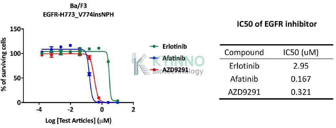
Ba/F3 cell, a murine interleukin-3 dependent pro-B cell line, is a popular system for exploring both kinases and their inhibitors, because some protein kinases can render the Ba/F3 cells to be depended on the activation of the kinases instead of IL-3 supplement, while their inhibitors can antagonize the kinase-dependent growth effects.

Specifications

Catalog NumberKC-0124
Cell Line NameBa/F3-EGFR-H773-V774ins_NPH Cell Line
Host Cell LineBa/F3
DescriptionStable Ba/F3 clone expressing exogenous EGFR gene bearing exon20 H773-V774 NPH insertion.
QuantityTwo vials of frozen cells (≥2-106/vial)
StabilityStable in culture over a minimum of 10 passages
ApplicationDrug screening and biological assays
Freezing Medium70% RPMI1640+20% FBS+10% DMSO
Propagation MediumRPMI1640+10%FBS
Selection MarkerPuromycin
MorphologyMostly single, round (some polymorph) cells in suspension
SubcultureSplit saturated culture 1:10 every 3 days; seed out at about 1-3 × 10⁵ cells/mL
Incubation37 °C with 5% CO2
StorageLiquid nitrogen immediately upon receiving
Doubling TimeApproximately 20 hours
Mycoplasma StatusNegative

Cell Line Generation

Characterization

Figure 1: Characterization of EGFR overexpression in the Ba/F3-EGFR-H773-V774ins_NPH stable clone using FACS.

Figure 2: Characterization of EGFR mutation in the Ba/F3-EGFR-H773-V774ins_NPH stable clone using PCR sequencing

Figure 3: Characterization of dose-response curves for EGFR inhibitors on Ba/F3-EGFR-H773-V774ins_NPH cells.

Cell Resuscitation

1. Prewarm culture medium (RPMI1640 + 10% FBS)in a 37°C water bath.
2. Thaw the frozen vial in a 37°C water bath for 1-2 minutes.
3. Transfer the vial into biosafety cabinet, and wipe the surface with 70% ethanol.
4. Unscrew the top of the vial and transfer the cell suspension gently into a sterile centrifuge tube containing 9.0mL complete culture medium.
5. Spin at ~ 125 × g for 5-7 minutes at room temperature, and discard the supernatant without disturbing the pellet.
6. Resuspend cell pellet with the appropriate volume of complete medium and transfer the cell suspension into a T25 culture flask.
7. Incubate the flask at 37°C, 5% CO2 incubator.
8. Split saturated culture 1:10 every 3 days; seed out at about 1-3 × 10⁵ cells/mL.

Cell Freezing

1. Prepare the freezing medium (70% RPMI-1640 + 20% FBS + 10% DMSO) fresh immediately before use.
2. Keep the freezing medium on ice and label cryovials.
3. Transfer cells to a sterile, conical centrifuge tube, and count the cells.
4. Centrifuge the cells at 250×g for 5 minutes at room temperature and carefully aspirate off the medium.
5. Resuspend the cells at a density of at least 3×10⁶ cells/mL in chilled freezing medium.
6. Aliquot 1 mL of the cell suspension into each cryovial.
7. Freeze cells in the CoolCell freezing container overnight in a -80°C freezer.
8. Transfer vials to liquid nitrogen for long-term storage.

References

1. Arcila, M. E. et al. EGFR Exon 20 Insertion Mutations in Lung Adenocarcinomas: Prevalence, Molecular Heterogeneity, and Clinicopathologic Characteristics. Molecular Cancer Therapeutics 12, 220–229 (2013)
Please enable JavaScript in your browser to complete this form.