MSLN (Mesothelin) is a cell-surface glycoprotein with normal expression in peritoneum, pleura, and pericardium, but with overexpression in a variety of cancers, including mesothelioma, pancreatic, lung, gastric, and ovarian cancers. It encodes a preproprotein that is proteolytically processed to generate two protein products, megakaryocyte potentiating factor and mesothelin. Mesothelin (MSLN) is an attractive antigen for chimeric antigen receptor (CAR) T therapy and the epitope selection within MSLN is essential. MSLN is an ideal cancer antigen for targeted immunotherapy. Diseases associated with MSLN include Benign Mesothelioma and Malignant Pleural Mesothelioma.
Specifications
Catalog Number
KC-5521
Cell Line Name
MIApaca2-MSLN Cell Line
Clone Number
3#
Host Cell Line
MIApaca2
Description
Stable MIApaca2 cell line expressing exogenous human MSLN gene
Quantity
One vial of frozen cells (≥2-106/vial)
Stability
Stable in culture over a minimum of 10 passages
Application
Drug screening and biological assays
Freezing Medium
70% RPMI1640 + 20% FBS + 10% DMSO
Propagation Medium
RPMI1640 + 10% FBS + 1μg/ml Puromycin
Selection Marker
Puromycin
Morphology
Epithelial
Subculture
Split saturated culture 1:2-1:3 every 2-3 days; seed out at about 1-2 × 10⁵ cells/ml
Incubation
37 °C with 5% CO2
Storage
Liquid nitrogen immediately upon receiving
Doubling Time
Approximately 30 hours
Mycoplasma Status
Negative
Cell Line Generation
MIApaca2-MSLN Cell Line was generated using a lentiviral vector expressing the human MSLN sequence.
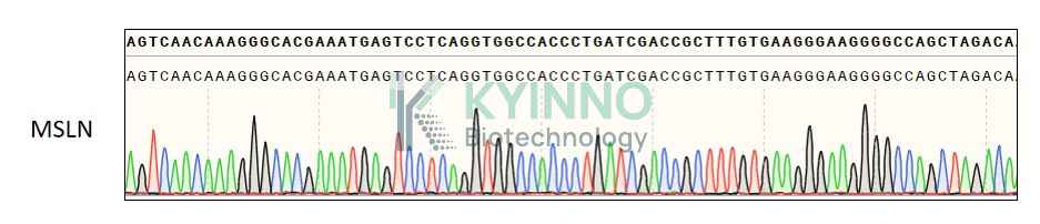
Characterization
Figure 1: Characterization of human MSLN overexpression in the MIApaca2-MSLN stable clone using PCR sequence
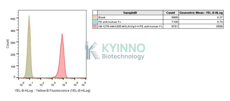
Figure 2: Characterization of human MSLN overexpression in the MIApaca2-MSLN stable clone using FACS.
Cell Resuscitation
1. Prewarm culture medium (RPMI1640 + 10% FBS + 1μg/ml Puromycin)in a 37°C water bath.
2. Thaw the frozen vial in a 37°C water bath for 1-2 minutes.
3. Transfer the vial into biosafety cabinet, and wipe the surface with 70% ethanol.
4. Unscrew the top of the vial and transfer the cell suspension gently into a sterile centrifuge tube containing 9.0mL complete culture medium.
5. Spin at ~ 125 × g for 5-7 minutes at room temperature, and discard the supernatant without disturbing the pellet.
6. Resuspend cell pellet with the appropriate volume of complete medium and transfer the cell suspension into a T25 culture flask.
7. Incubate the flask at 37°C, 5% CO2 incubator.
8. Split saturated culture 1:2-1:3 every 2-3 days; seed out at about 1-2 × 10⁵ cells/mL.
Cell Freezing
1. Prepare the freezing medium (70% RPMI1640 + 20% FBS + 10% DMSO) fresh immediately before use.
2. Keep the freezing medium on ice and label cryovials.
3. Transfer cells to a sterile, conical centrifuge tube, and count the cells.
4. Centrifuge the cells at 250×g for 5 minutes at room temperature and carefully aspirate off the medium.
5. Resuspend the cells at a density of at least 3×10⁶ cells/mL in chilled freezing medium.
6. Aliquot 1 mL of the cell suspension into each cryovial.
7. Freeze cells in the CoolCell freezing container overnight in a -80°C freezer.
8. Transfer vials to liquid nitrogen for long-term storage.
References
1. Zhang, Z., Jiang, D., Yang, H. et al. Modified CAR T cells targeting membrane-proximal epitope of mesothelin enhances the antitumor function against large solid tumor. Cell Death Dis 10, 476 (2019).10.
2. Li Y, Tian W, Zhang H, Zhang Z, Zhao Q, Chang L, Lei N, Zhang W. MSLN Correlates With Immune Infiltration and Chemoresistance as a Prognostic Biomarker in Ovarian Cancer. Front Oncol. 2022 May 25;12:830570.
Use License Agreement
Research Use Only.
Not for use in diagnostic procedures or therapeutic applications.
Redistribution of the cell line or its derivatives is prohibited without prior written permission from Kyinno Biotechnology.