KC-4837

CT26-ERBB2-GFP-Luc2-Cell-Line

×
Please enable JavaScript in your browser to complete this form.
56707
Home » CT26-ERBB2-GFP-Luc2-Cell-Line

Background of CT26-ERBB2-GFP-Luc2-Cell-Line

ERBB2, also known as CD340, encodes a member of the epidermal growth factor (EGF) receptor tyrosine kinase family. This protein itself does not have a ligand binding domain, so it cannot bind to growth factors. However, it does tightly bind with other ligands in the EGF receptor family to form heterodimers, stabilizing ligand binding and enhancing activation of downstream signaling pathways mediated by kinases, such as those involving mitogen activated protein kinase and phosphatidylinositol 3-kinase. It is reported that the amplification and/or overexpression of this gene are found in many cancers, including breast cancer and ovarian tumors.
Luciferase is an oxidative enzyme that can produce bioluminescence with addition of luciferin, but doesn’t need an external light source unlike fluorescent proteins. Photo emission can be detected directly by light sensitive devices. Such as luminometers or modified microscopes. Luciferase is widely used in many fields of biological research, such as transcriptional activity, kinase or other enzyme activity, cellular ATP level, and whole animal imaging.

Specifications

Catalog NumberKC-4837
Cell Line NameCT26-ERBB2-GFP-Luc2-Cell-Line
Clone Number26-8#
Host Cell LineCT26
DescriptionStable CT26 clone expressing exogenous ERBB2 and GFP and Luciferase gene.
QuantityTwo vials of frozen cells (≥2-106/vial)
StabilityStable in culture over a minimum of 10 passages
ApplicationDrug screening and biological assays
Freezing Medium70%RPMI 1640 + 20% FBS + 10% DMSO
Propagation MediumRPMI 1640 + 10% FBS
Selection MarkerNA
MorphologyFibroblast
SubcultureSplit saturated culture 1:4-1:10 every 2-3 days; seed out at about 1-3 × 10⁵ cells/mL
Incubation37 °C with 5% CO2
StorageLiquid nitrogen immediately upon receiving
Doubling TimeApproximately 24 hours
Mycoplasma StatusNegative

Cell Line Generation

CT26-ERBB2-GFP-Luc2-cell-line was generated using a lentiviral vector expressing the ERBB2-GFP-Luc2 sequence.

Characterization

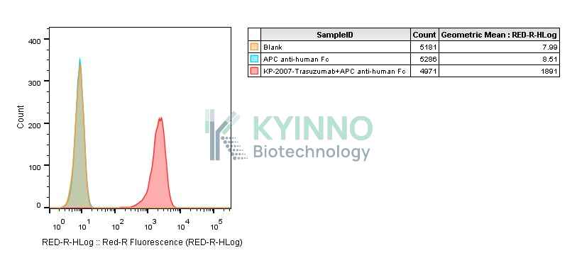
Figure 1: Characterization of ERBB2 overexpression in the CT26-ERBB2-GFP-Luc2 stable clone using FACS.

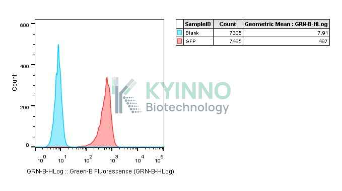
Figure 2: Characterization of GFP overexpression in the CT26-ERBB2-GFP-Luc2 stable clone using FACS.

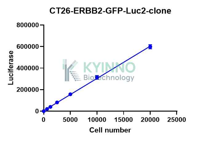
Figure 3: Characterization of the CT26-ERBB2-GFP-Luc2-cell-line stable clone using Bright-Lite Luciferase Assay System in the conditions of different cell number.

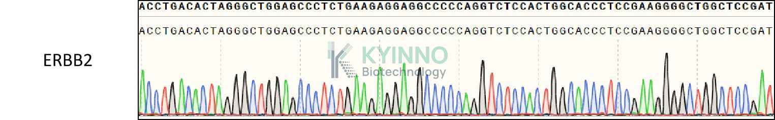
Figure 4: Characterization of ERBB2 in the CT26-ERBB2-GFP-Luc2 stable clone using PCR sequencing.

Cell Resuscitation

1. Prewarm culture medium (RPMI 1640 supplemented with 10% FBS)in a 37°C water bath.
2. Thaw the frozen vial in a 37°C water bath for 1-2 minutes.
3. Transfer the vial into biosafety cabinet, and wipe the surface with 70% ethanol.
4. Unscrew the top of the vial and transfer the cell suspension gently into a sterile centrifuge tube containing 9.0mL complete culture medium.
5. Spin at ~ 125 × g for 5-7 minutes at room temperature, and discard the supernatant without disturbing the pellet.
6. Resuspend cell pellet with the appropriate volume of complete medium and transfer the cell suspension into a T25 culture flask.
7. Incubate the flask at 37°C, 5% CO2 incubator.
8. Split saturated culture 1:4-1:10 every 2-3 days; seed out at about 1-3 × 10⁵ cells/mL.

Cell Freezing

1. Prepare the freezing medium (70%RPMI 1640 + 20% FBS + 10% DMSO) fresh immediately before use.
2. Keep the freezing medium on ice and label cryovials.
3. Transfer cells to a sterile, conical centrifuge tube, and count the cells.
4. Centrifuge the cells at 250×g for 5 minutes at room temperature and carefully aspirate off the medium.
5. Resuspend the cells at a density of at least 3×10⁶ cells/mL in chilled freezing medium.
6. Aliquot 1 mL of the cell suspension into each cryovial.
7. Freeze cells in the CoolCell freezing container overnight in a -80°C freezer.
8. Transfer vials to liquid nitrogen for long-term storage.

References

1.Greer LF, Szalay AA (2002). Imaging of light emission from the expression of luciferases in living cells and organisms: a review. Luminescence. 17 (1): 43–74. doi:10.1002/bio.676. PMID 11816060.
2.Idamakanti M, Bijjam RI, Kumar M, Mukkamalla SK. Long Durable Response With Trastuzumab Deruxtecan Monotherapy in a Triple-Negative Metastatic Breast Cancer Patient With Human Epidermal Growth Factor Receptor 2 Mutation: A Long-Term Follow-Up and Literature Review. J Med Cases. 2025 Jun 16;16(6):212-221. doi: 10.14740/jmc5136. PMID: 40642740; PMCID: PMC12239829.
3.Zheng Y, Shen G, Zhang C, Huo X, Xin Y, Fang Q, Guan Y, Zhao F, Ren D, Liu Z, Wang M, Zhao J. Efficacy of anti-HER2 drugs in the treatment of patients with HER2-mutated cancers: a systematic review and meta-analysis. Clin Exp Med. 2023 Nov;23(7):3205-3216. doi: 10.1007/s10238-023-01072-7. Epub 2023 Apr 30. PMID: 37120775.
Please enable JavaScript in your browser to complete this form.