kc-5758

293T-STAT5-Luc2-IL2RA-IL2RB-IL2RG-PD1-Middle Cell Line

×
Please enable JavaScript in your browser to complete this form.
58986
Home » 293T-STAT5-Luc2-IL2RA-IL2RB-IL2RG-PD1-Middle Cell Line

Background of 293T-STAT5-Luc2-IL2RA-IL2RB-IL2RG-PD1-Middle Cell Line

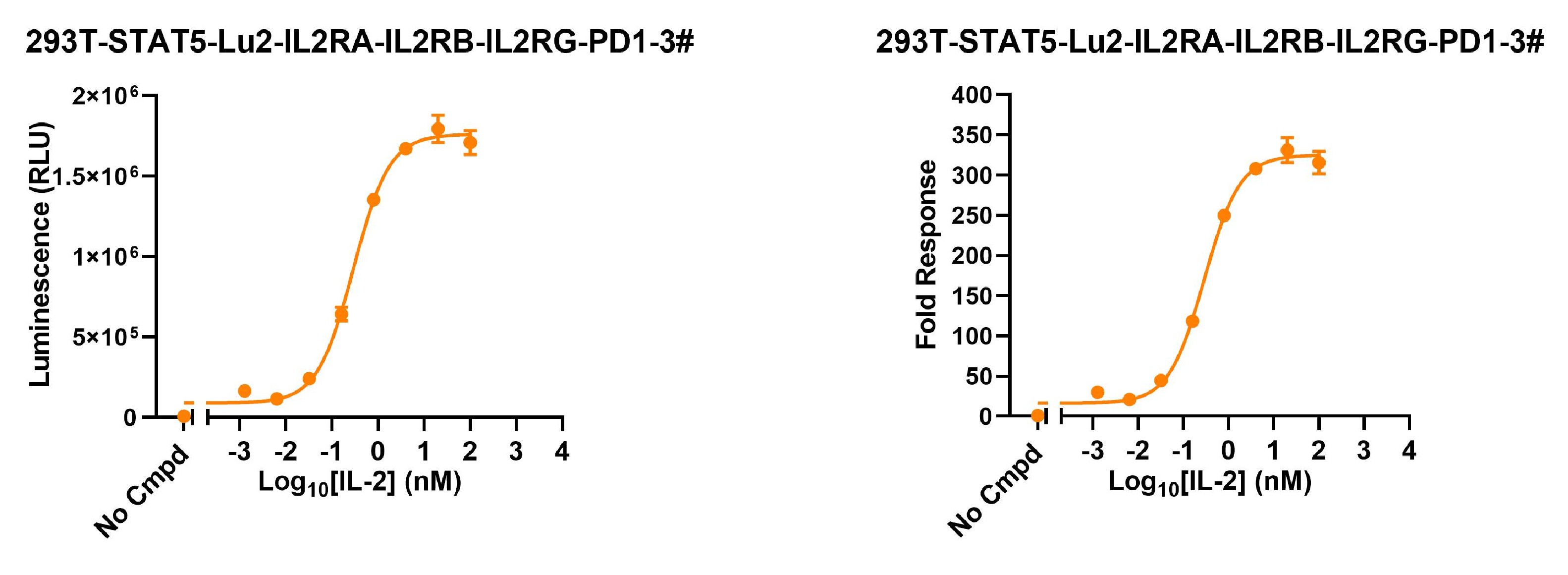
The protein encoded by STAT5 is a member of the STAT family of transcription factors. In response to cytokines and growth factors, STAT family members are phosphorylated by the receptor associated kinases, and then form homo- or heterodimers that translocate to the cell nucleus where they act as transcription activators. STAT5 is activated by, and mediates the responses of many cell ligands, such as IL2, IL3, IL7 GM-CSF, erythropoietin, thrombopoietin, and different growth hormones. Activation of STAT5 in myeloma and lymphoma associated with a TEL/JAK2 gene fusion is independent of cell stimulus and has been shown to be essential for tumorigenesis.STAT5 plays an important role in the maintenance of normal immune function and homeostasis, both of which are regulated by specific members of IL-2 family of cytokines, which share a common gamma chain (γc) in their receptor complex. STAT5 critically mediates the biological actions of members of the γc family of cytokines in the immune system. 
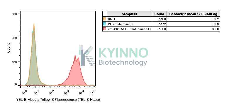
PD-1 is a transmembrane glycoprotein expressed on T, B, and Dentric cells. This molecule functions as a checkpoint in T cell proliferation. Ligation of PD-1 with its ligands inhibits the production of IL-2, IL-7, IL-10, and IL-12 as well as other cytokines by macrophages, natural killer (NK) cells, and T cells, which can suppress cell proliferation and inflammation.

Specifications

Catalog Numberkc-5758
Cell Line Name293T-STAT5-Luc2-IL2RA-IL2RB-IL2RG-PD1-Middle Cell Line
Clone Number3#
Host Cell Line293T
DescriptionStable 293T clone expressing STAT5 reporter gene and human IL-2 receptor (IL2RA/B/G) co-expressed with PD1
QuantityTwo vials of frozen cells (≥2-106/vial)
StabilityStable in culture over a minimum of 10 passages
ApplicationDrug screening and biological assays
Freezing Medium70% DMEM + 20% FBS + 10% DMSO
Propagation MediumDMEM+10% FBS+0.5 μg/mL puromycin+75μg/mL Hygromycin B+50 μg/mL Zeocin
Selection MarkerPuromycin | Hygromycin B | Zeocin
MorphologyEpithelial
SubcultureSplit saturated culture 1:4-1:8 every 2-3 days; seed out at about 1-3 × 10⁵ cells/mL
Incubation37 °C with 5% CO2
StorageLiquid nitrogen immediately upon receiving
Doubling TimeApproximately 30 hours
Mycoplasma StatusNegative

Cell Line Generation

293T-STAT5-Luc2-IL2RA-IL2RB-IL2RG-PD1 cell line was generated using lentivirus expressing IL2RA&IL2RB&IL2RG&PD1 sequence.

Characterization

Figure1: 293T-STAT5-Luc2-IL2RA-IL2RB-IL2RG-PD1 cell line was seeded into the 96-well plate, and treated with IL2 at a maximum concentration 100nM for 16 hours and readout with Bright-Glo system.

Figure 2: Characterization of PD1 overexpression in the 293T-STAT5-Luc2-IL2RA-IL2RB-IL2RG-PD1 stable clone using FACS.

Figure 3: Characterization of IL2RA&IL2RB&IL2RG&PD1 in the 293T-STAT5-Luc2-IL2RA-IL2RB-IL2RG-PD1 stable clone using PCR sequencing.

Cell Resuscitation

  1. Prewarm culture medium (DMEM + 10% FBS + 0.5μg/mL Puromycin + 75µg/mL Hygromycin B + 50µg/mL zeocin) in a 37°C water bath.
  2. Thaw the frozen vial in a 37°C water bath for 1-2 minutes.
  3. Transfer the vial into biosafety cabinet, and wipe the surface with 70% ethanol.
  4. Unscrew the top of the vial and transfer the cell suspension gently into a sterile centrifuge tube containing 9.0mL complete culture medium.
  5. Spin at ~ 125 × g for 5-7 minutes at room temperature, and discard the supernatant without disturbing the pellet.
  6. Resuspend cell pellet with the appropriate volume of complete medium and transfer the cell suspension into a T25 culture flask.
  7. Incubate the flask at 37°C, 5% CO2 incubator.
  8. Split saturated culture 1:4-1:8 every 2-3 days; seed out at about 1-3 × 10⁵ cells/mL.

Cell Freezing

  1. Prepare the freezing medium (70% DMEM + 20% FBS + 10% DMSO) fresh immediately before use.
  2. Keep the freezing medium on ice and label cryovials.
  3. Transfer cells to a sterile, conical centrifuge tube, and count the cells.
  4. Centrifuge the cells at 250×g for 5 minutes at room temperature and carefully aspirate off the medium.
  5. Resuspend the cells at a density of at least 3×10⁶ cells/mL in chilled freezing medium.
  6. Aliquot 1 mL of the cell suspension into each cryovial.
  7. Freeze cells in the CoolCell freezing container overnight in a -80°C freezer.
  8. Transfer vials to liquid nitrogen for long-term storage.

References

  1. Rani A, Murphy JJ. STAT5 in Cancer and Immunity. J Interferon Cytokine Res. 2016 Apr;36(4):226-37. doi: 10.1089/jir.2015.0054. Epub 2015 Dec 30. PMID: 26716518.
  2. Villarino AV, Laurence AD, Davis FP, Nivelo L, Brooks SR, Sun HW, Jiang K, Afzali B, Frasca D, Hennighausen L, Kanno Y, O'Shea JJ. A central role for STAT5 in the transcriptional programing of T helper cell metabolism. Sci Immunol. 2022 Nov 25;7(77):eabl9467. doi: 10.1126/sciimmunol.abl9467. Epub 2022 Nov 25. PMID: 36427325; PMCID: PMC9844264.
  3. Kim U, Shin HY. Genomic Mutations of the STAT5 Transcription Factor Are Associated with Human Cancer and Immune Diseases. Int J Mol Sci. 2022 Sep 25;23(19):11297. doi: 10.3390/ijms231911297. PMID: 36232600; PMCID: PMC9569778.
  4. Rezayi M, Hosseini A. Structure of PD1 and its mechanism in the treatment of autoimmune diseases. Cell Biochem Funct. 2023 Oct;41(7):726-737. doi: 10.1002/cbf.3827. Epub 2023 Jul 20. PMID: 37475518.
Please enable JavaScript in your browser to complete this form.