KC-6138

22RV1-CHD7-KO-mcherry-Luc2 Cell Line

×
Please enable JavaScript in your browser to complete this form.
60818
Home » 22RV1-CHD7-KO-mcherry-Luc2 Cell Line

Background of 22RV1-CHD7-KO-mcherry-Luc2 Cell Line

Luciferase is an oxidative enzyme that can produce bioluminescence with the adddition of luciferin,but don’t need an external light source,which is different from fluorescent proteins.The bioluminescence can be detected directly by light sensitive device,such as luminometer or modified microscope.Luciferase is widely used in many fields ofbiological research,such as transcriptional activity,kinase or other enzyme activity,cellular ATP level,whole animal imaging.

Specifications

Catalog NumberKC-6138
Cell Line Name22RV1-CHD7-KO-mcherry-Luc2 Cell Line
NCBI/UniProt Accession NumberN/A
Clone Number14#
Host Cell Line22RV1-CHD7-KO
DescriptionStable 22RV1-CHD7-KO clone with mcherry and Luciferase gene overexpression
QuantityTwo vials of frozen cells (≥2-106/vial)
StabilityStable in culture over a minimum of 10 passages
ApplicationDrug screening and biological assays
Freezing MediumRPMI1640+20% FBS+10% DMSO
Propagation MediumRPMI1640+10% FBS+300μg/mL Hygromycin B
Selection MarkerHygromycin B
MorphologyEpithelial
SubcultureSplit saturated culture 1:2 every 2-3 days; seed out at about 1-3 × 10⁵ cells/mL
Incubation37 °C with 5% CO2
StorageLiquid nitrogen immediately upon receiving
Doubling TimeApproximately 30 hours
Mycoplasma StatusNegative

Cell Line Generation

22RV1-CHD7-KO-mcherry-Luc2 Cell Line was generated using a lentiviral vector expressing mcherry and luciferase sequence.

Characterization

Figure 1: Characterization of 22Rv1-CHD7-KO cell line stable clone using PCR sequencing

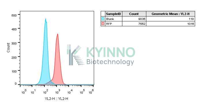
Figure 2: Characterization of mcherry overexpression in the 22RV1-CHD7-KO-mcherry-Luc2 stable clone using FACS.

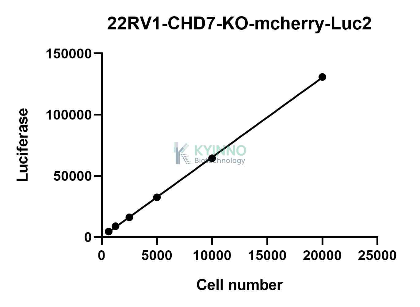
Figure 3: Characterization of luciferase expression in 22RV1-CHD7-KO-mcherry-Luc2 cell using the Bright-Glo Detection.

Cell Resuscitation

  1. Prewarm culture medium (RPMI1640 + 10% FBS + 300μg/mL Hygromycin B) in a 37°C water bath.
  2. Thaw the frozen vial in a 37°C water bath for 1-2 minutes.
  3. Transfer the vial into biosafety cabinet, and wipe the surface with 70% ethanol.
  4. Unscrew the top of the vial and transfer the cell suspension gently into a sterile centrifuge tube containing 9.0mL complete culture medium.
  5. Spin at ~ 125 × g for 5-7 minutes at room temperature, and discard the supernatant without disturbing the pellet.
  6. Resuspend cell pellet with the appropriate volume of complete medium and transfer the cell suspension into a T25 culture flask.
  7. Incubate the flask at 37°C, 5% CO2 incubator.
  8. Split saturated culture 1:2 every 2-3 days; seed out at about 1-3 × 10⁵ cells/mL.

Cell Freezing

  1. Prepare the freezing medium (70% RPMI1640 + 20% FBS + 10% DMSO) fresh immediately before use.
  2. Keep the freezing medium on ice and label cryovials.
  3. Transfer cells to a sterile, conical centrifuge tube, and count the cells.
  4. Centrifuge the cells at 250×g for 5 minutes at room temperature and carefully aspirate off the medium.
  5. Resuspend the cells at a density of at least 3×10⁶ cells/mL in chilled freezing medium.
  6. Aliquot 1 mL of the cell suspension into each cryovial.
  7. Freeze cells in the CoolCell freezing container overnight in a -80°C freezer.
  8. Transfer vials to liquid nitrogen for long-term storage.

References

  1. Greer LF 3rd,Szalay AA.Imaging of light emission from the expression of luciferases in living cells and organisms:a review.Luminescence.2002 Jan-Feb;17(1):43-74.doi:10.1002/bio.676.PMID:11816060.
Please enable JavaScript in your browser to complete this form.