KC-6049

MCF7-ESR1-G521S-Y537S-KI Cell Line

×
Please enable JavaScript in your browser to complete this form.
62516
Home » MCF7-ESR1-G521S-Y537S-KI Cell Line

Background of MCF7-ESR1-G521S-Y537S-KI Cell Line

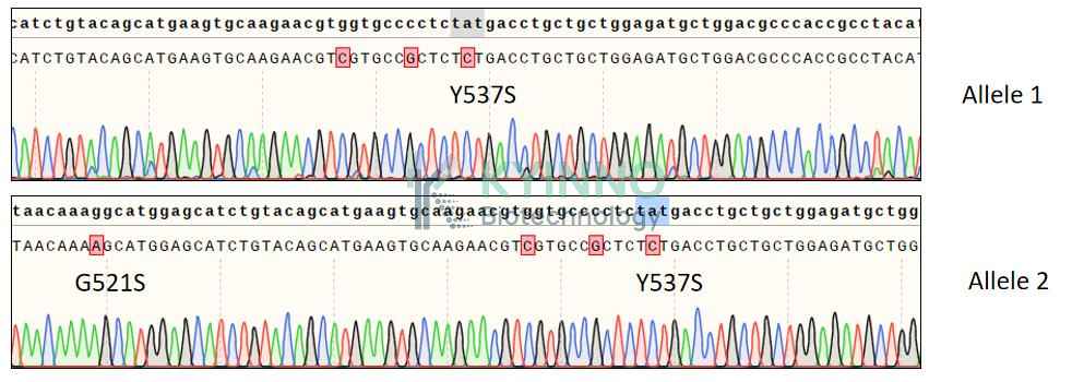
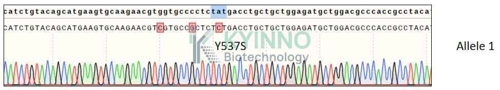
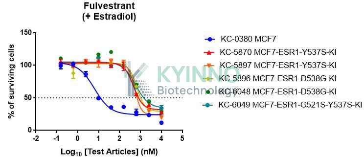
ESR1-G521S-Y537S is a complex, double mutation in the estrogen receptor alpha (ERα) gene, combining a glycine-to-serine substitution at codon 521 within the ligand-binding domain (LBD) with the well-characterized activating Y537S mutation. This dual alteration represents a rare but clinically significant variant associated with profound endocrine therapy resistance in hormone receptor-positive breast cancer. The G521S mutation, located near the helix 11-12 region, may synergize with Y537S to further stabilize the active conformation of ERα, enhance constitutive transcriptional activity, and alter coregulator recruitment beyond that observed with single mutations. Mechanistically, the double mutation confers resistance to a broader spectrum of endocrine agents, including selective estrogen receptor degraders (SERDs) like fulvestrant and next-generation oral SERDs, while potentially retaining sensitivity to selective estrogen receptor covalent antagonists (SERCAs) and proteolysis-targeting chimeras (PROTACs). Detection of complex ESR1 mutations, including G521S-Y537S, in circulating tumor DNA (ctDNA) is associated with poor prognosis, rapid disease progression, and limited response to conventional endocrine therapies. These compound mutations often arise under sequential endocrine treatment pressure through clonal evolution and represent a formidable challenge in precision oncology. Understanding the structural and functional consequences of such double mutations is critical for developing effective therapeutic strategies for heavily pretreated, endocrine-resistant breast cancer.

Specifications

Catalog NumberKC-6049
Cell Line NameMCF7-ESR1-G521S-Y537S-KI Cell Line
Clone Number1A6
Host Cell LineMCF7
DescriptionStable MCF7 clone expressing endogenous ESR1 gene bearing G521S and Y537S mutations.
QuantityTwo vials of frozen cells (≥2-106/vial)
StabilityStable in culture over a minimum of 10 passages
ApplicationDrug screening and biological assays
Freezing MediumMEM+20% FBS+10% DMSO
Propagation MediumMEM+10% FBS+0.01mM NEAA+0.01mg/ml insulin
Selection MarkerNA
MorphologyEpithelial
SubcultureSplit saturated culture 1:2-1:4 every 2-3 days; seed out at about 1-3 × 10⁵ cells/mL
Incubation37 °C with 5% CO2
StorageLiquid nitrogen immediately upon receiving
Doubling TimeApproximately 30 hours
Mycoplasma StatusNegative

Cell Line Generation

MCF7-ESR1-G521S-Y537S-KI cell line was generated using the CRISPR method.

Characterization

Figure 1: Characterization of MCF7-ESR1-G521S-Y537S-KI cell line stable clone using PCR sequencing.

Figure 2: Characterization of MCF7-ESR1-G521S-Y537S-KI cell line stable clone using RT-PCR sequencing.

Figure 3. Characterization of dose-response curves for ESR1 inhibitors on MCF7 and MCF7-ESR1-G521S-Y537S-KI cells.

Cell Resuscitation

1. Prewarm culture medium (MEM+10% FBS+0.01mM NEAA+0.01mg/ml insulin)in a 37°C water bath.
2. Thaw the frozen vial in a 37°C water bath for 1-2 minutes.
3. Transfer the vial into biosafety cabinet, and wipe the surface with 70% ethanol.
4. Unscrew the top of the vial and transfer the cell suspension gently into a sterile centrifuge tube containing 9.0mL complete culture medium.
5. Spin at ~ 125 × g for 5-7 minutes at room temperature, and discard the supernatant without disturbing the pellet.
6. Resuspend cell pellet with the appropriate volume of complete medium and transfer the cell suspension into a T25 culture flask.
7. Incubate the flask at 37°C, 5% CO2 incubator.
8. Split saturated culture 1:2-1:4 every 2-3 days; seed out at about 1-3 × 10⁵ cells/mL

Cell Freezing

  1. Prepare the freezing medium (MEM+20% FBS+10% DMSO) fresh immediately before use.
  2. Keep the freezing medium on ice and label cryovials.
  3. Transfer cells to a sterile, conical centrifuge tube, and count the cells.
  4. Centrifuge the cells at 250×g for 5 minutes at room temperature and carefully aspirate off the medium.
  5. Resuspend the cells at a density of at least 3×10⁶ cells/mL in chilled freezing medium.
  6. Aliquot 1 mL of the cell suspension into each cryovial.
  7. Freeze cells in the CoolCell freezing container overnight in a -80°C freezer.
  8. Transfer vials to liquid nitrogen for long-term storage

References

  1. Toy, W., et al. (2017). "Activating ESR1 mutations and resistance to endocrine therapy in breast cancer." Nature Reviews Cancer, 17(7), 424-435.
  2. Zoppoli, G., et al. (2024). "Compound ESR1 mutations in metastatic breast cancer: mechanisms and clinical implications." Cancer Discovery, 14(3), 456-471.
  3. Brett, J.O., et al. (2025). "Next-generation endocrine therapies for ESR1-mutant breast cancer." Clinical Cancer Research, 31(2), 245-258.
Please enable JavaScript in your browser to complete this form.