KC-6161

293T-STAT5-Luc2-IL2RB-IL2RG-PD1-Middle Cell Line

×
请在浏览器中启用JavaScript来完成此表单。
63281
Home » 293T-STAT5-Luc2-IL2RB-IL2RG-PD1-Middle Cell Line

Background of 293T-STAT5-Luc2-IL2RB-IL2RG-PD1-Middle Cell Line

The protein encoded by STAT5 is a member of the STAT family of transcription factors. In response to cytokines and growth factors, STAT family members are phosphorylated by the receptor associated kinases, and then form homo- or heterodimers that translocate to the cell nucleus where they act as transcription activators. STAT5 is activated by, and mediates the responses of many cell ligands, such as IL2, IL3, IL7 GM-CSF, erythropoietin, thrombopoietin, and different growth hormones. Activation of STAT5 in myeloma and lymphoma associated with a TEL/JAK2 gene fusion is independent of cell stimulus and has been shown to be essential for tumorigenesis.STAT5 plays an important role in the maintenance of normal immune function and homeostasis, both of which are regulated by specific members of IL-2 family of cytokines, which share a common gamma chain (γc) in their receptor complex. STAT5 critically mediates the biological actions of members of the γc family of cytokines in the immune system. PD-1 is a transmembrane glycoprotein expressed on T, B, and Dentric cells. This molecule functions as a checkpoint in T cell proliferation. Ligation of PD-1 with its ligands inhibits the production of IL-2, IL-7, IL-10, and IL-12 as well as other cytokines by macrophages, natural killer (NK) cells, and T cells, which can suppress cell proliferation and inflammation.

Specifications

Catalog NumberKC-6161
Cell Line Name293T-STAT5-Luc2-IL2RB-IL2RG-PD1-Middle Cell Line
NCBI/UniProt Accession NumberQ15116
Clone Number24#
Host Cell Line293T-STAT5-Luc2-IL2RB-IL2RG
DescriptionStable 293T-STAT5-Luc2-IL2RB-IL2RG cell line expressing exogenous PD1 gene
QuantityOne vial of frozen cells (≥2-106/vial)
StabilityStable in culture over a minimum of 10 passages
ApplicationDrug screening and biological assays
Freezing Medium70% basal medium+20% FBS+10% DMSO
Propagation MediumDMEM+10%FBS+50µg/mL Zeocin+75µg/mL hygromycin B+0.5µg/mL puromycin
Selection Markerhygromycin B, Zeocin, puromycin
MorphologyEpithelial-like
SubcultureSplit saturated culture 1:4-1:8 every 2-3 days
Incubation37 °C with 5% CO2
StorageLiquid nitrogen immediately upon receiving
Doubling TimeApproximately 30 hours
Mycoplasma StatusNegative
In Vivo ValidationNA

Cell Line Generation

293T-STAT5-Luc2-IL2RB-IL2RG-PD1-Middle cell line was generated using a lentiviral vector expressing the PD1 sequence in Middle level.

Characterization

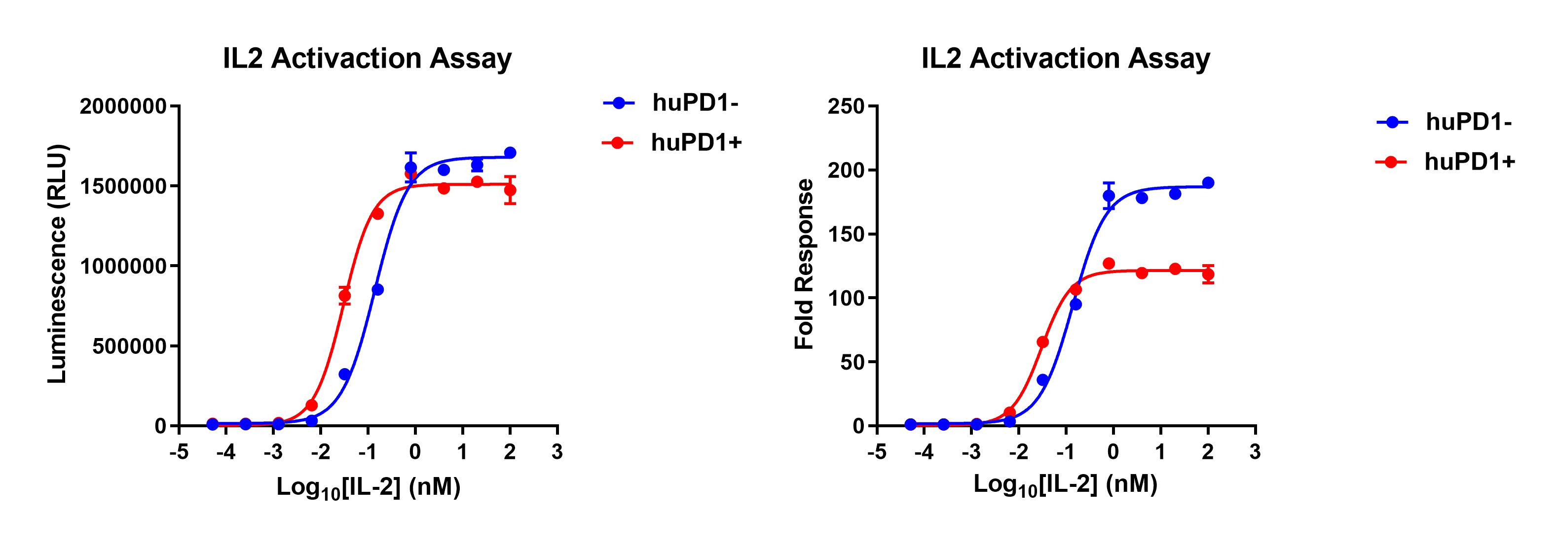
Figure 1. 293T-STAT5-Luc2-IL2RB-IL2RG-PD1-Middle cell line was seeded into the 96-well plate, and treated with IL-2 at a maximum concentration of 100nM for 16 hours, then readout with Bright-Glo system.

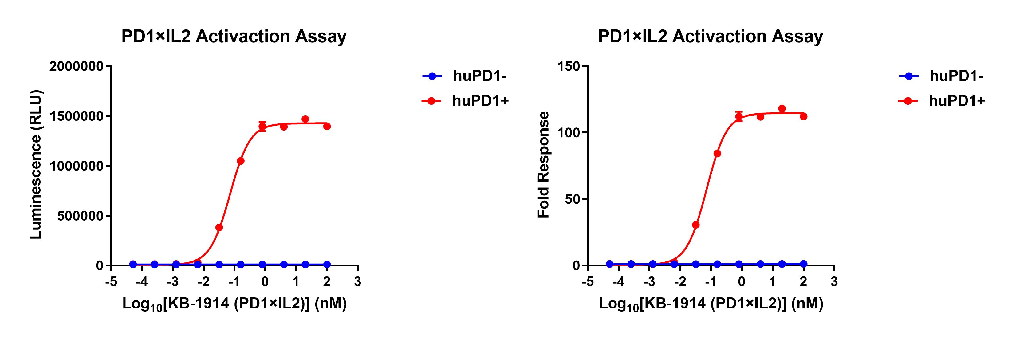
Figure 2. 293T-STAT5-Luc2-IL2RB-IL2RG-PD1-Middle cell line was seeded into the 96-well plate, and treated with KB-1914(PD1×IL2) at a maximum concentration of 100nM for 16 hours, then readout with Bright-Glo system.

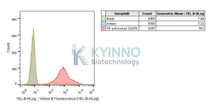
Figure 3. Characterization of PD1 overexpression in the 293T-STAT5-Luc2-IL2RB-IL2RG-PD1-Middle stable clone using FACS.

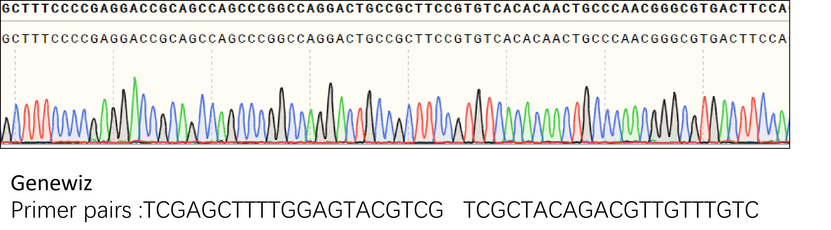
Figure 4. Characterization of PD1 in the 293T-STAT5-Luc2-IL2RB-IL2RG-PD1-Middle stable clone using PCR sequencing.

Cell Resuscitation

  1. Pre-warm complete culture medium (basal medium and 10% FBS) in a 37°C water bath.
  2. Rapidly thaw the cryovial in a 37°C water bath for 1-2 minutes with gentle agitation.
  3. Transfer the vial to a biosafety cabinet, and disinfect the exterior with 70% ethanol.
  4. Aseptically transfer the cell suspension dropwise into a sterile centrifuge tube containing 9.0 mL of pre-warmed complete medium.
  5. Centrifuge at approximately 125 × g for 5–7 minutes at room temperature, carefully aspirate the supernatant without disturbing the cell pellet.
  6. Gently resuspend the pellet in an appropriate volume of complete medium and transfer the suspension into a T25 flask.
  7. Incubate the flask in a 37°C in a humidified 5% CO2 incubator.
  8. Assess cell viability and morphology after 24 hours. If cells appear healthy, replace the medium with fresh medium supplemented with the appropriate selective antibiotic.
  9. Subculture the cells at a ratio of 1:4-1:8 every 2-3 days upon reaching 80%–90% confluency.

Cell Freezing

  1. Prepare the freezing medium (70% basal medium, 20% FBS and 10% DMSO) freshly before use.
  2. Pre-chill the freezing medium on ice and label the cryovials accordingly.
  3. Transfer the cell suspension to a sterile conical tube and perform a cell count to determine total viability and density.
  4. Centrifuge the cells at 250×g for 5 minutes at room temperature; carefully aspirate the supernatant.
  5. Gently resuspend the cell pellet in chilled freezing medium, ensuring a minimum cell density of 3×10⁶ cells/mL.
  6. Aliquot 1 mL of the cell suspension into each pre-labeled cryovial.
  7. Place the cryovials into a CoolCell® container and store at -80°C overnight for controlled-rate cooling.
  8. Transfer the cryovials to the liquid nitrogen for long-term storage the folMiddleing day.

References

1. Rani A, Murphy JJ. STAT5 in Cancer and Immunity. J Interferon Cytokine Res. 2016 Apr;36(4):226-37. doi: 10.1089/jir.2015.0054. Epub 2015 Dec 30. PMID: 26716518.
2. Villarino AV, Laurence AD, Davis FP, Nivelo L, Brooks SR, Sun HW, Jiang K, Afzali B, Frasca D, Hennighausen L, Kanno Y, O'Shea JJ. A central role for STAT5 in the transcriptional programing of T helper cell metabolism. Sci Immunol. 2022 Nov 25;7(77):eabl9467. doi: 10.1126/sciimmunol.abl9467. Epub 2022 Nov 25. PMID: 36427325; PMCID: PMC9844264.
3. Kim U, Shin HY. Genomic Mutations of the STAT5 Transcription Factor Are Associated with Human Cancer and Immune Diseases. Int J Mol Sci. 2022 Sep 25;23(19):11297. doi: 10.3390/ijms231911297. PMID: 36232600; PMCID: PMC9569778.
4. Rezayi M, Hosseini A. Structure of PD1 and its mechanism in the treatment of autoimmune diseases. Cell Biochem Funct. 2023 Oct;41(7):726-737. doi: 10.1002/cbf.3827. Epub 2023 Jul 20. PMID: 37475518.

Use License Agreement

Research Use Only.
Not for use in diagnostic procedures or therapeutic applications.
Redistribution of the cell line or its derivatives is prohibited without prior written permission from Kyinno Biotechnology.
请在浏览器中启用JavaScript来完成此表单。