KC-6310

293T-ARRB2-SmBiT-KI Cell Line

×
请在浏览器中启用JavaScript来完成此表单。
64096
Home » 293T-ARRB2-SmBiT-KI Cell Line

Background of 293T-ARRB2-SmBiT-KI Cell Line

The ARRB2 gene maps to chromosome 17p13.2 and encodes β-arrestin 2, a ubiquitously expressed multifunctional adaptor protein of the arrestin family. Also termed β-arrestin 2 or non-visual arrestin 2, ARRB2 acts as a core regulator of G protein-coupled receptor (GPCR) signaling. It mediates GPCR desensitization, internalization and downstream signal transduction, which fine-tunes cellular responses to extracellular stimuli and maintains physiological homeostasis by preventing excessive signal amplification. Beyond canonical GPCR regulation, ARRB2 is widely expressed in the central nervous system, immune cells and peripheral organs, and modulates cell proliferation, migration and survival across tissues. Aberrant expression of ARRB2 is linked to multiple disorders. In intrahepatic cholangiocarcinoma, elevated ARRB2 accelerates malignant progression and induces pemigatinib resistance via the MAPK and Hippo/YAP signaling pathways. ARRB2 exerts dual effects in glioblastoma and promotes tumor development in prostate cancer; it also alleviates inflammation and fibrosis in non-neoplastic diseases. Accordingly, ARRB2 is recognized as a valuable prognostic biomarker and therapeutic target. Small-molecule inhibitors and CRISPR-based tools have been developed to manipulate its activity for disease intervention. 
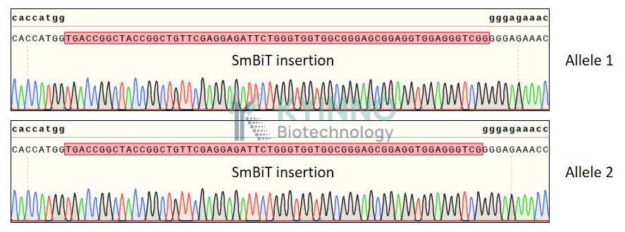
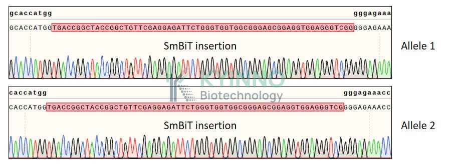
The ARRB2-SmBiT-KI model is a robust tool for analyzing endogenous ARRB2 dynamics. Using CRISPR-Cas9 knock-in technology, the 11-amino-acid SmBiT subunit of NanoLuc luciferase is fused to the endogenous ARRB2 locus. This system allows sensitive real-time detection of ARRB2 localization, trafficking and protein-protein interactions under physiological conditions, free from overexpression artifacts. It has become a key platform for drug discovery, functional genomics and mechanistic research on GPCR signaling.

Specifications

Catalog NumberKC-6310
Cell Line Name293T-ARRB2-SmBiT-KI Cell Line
NCBI/UniProt Accession NumberNM_004313.4/P32121
Clone Number3B1
Host Cell Line293T
Description293T cell line stable clone with endogenous SmBiT-tagged ARRB2 gene
QuantityTwo vials of frozen cells (≥2-106/vial)
StabilityStable in culture over a minimum of 10 passages
ApplicationDrug screening and biological assays
Freezing Medium70% DMEM + 20% FBS + 10% DMSO
Propagation MediumDMEM + 10% FBS
Selection MarkerNA
MorphologyEpithelial
SubcultureSplit saturated culture 1:4-1:8 every 2-3 days; seed out at about 1-3 × 10⁵ cells/mL
Incubation37 °C with 5% CO2
StorageLiquid nitrogen immediately upon receiving
Doubling TimeApproximately 30 hours
Mycoplasma StatusNegative

Cell Line Generation

293T-ARRB2-SmBiT-KI cell line was generated using the CRISPR method.

Characterization

Figure 1: Characterization of 293T-ARRB2-SmBiT-KI cell line stable clone using PCR sequencing.

Figure 2: Characterization of 293T-ARRB2-SmBiT-KI cell line stable clone using RT-PCR sequencing.

Cell Resuscitation

  1. Prewarm culture medium (DMEM + 10% FBS) in a 37°C water bath.
  2. Thaw the frozen vial in a 37°C water bath for 1-2 minutes.
  3. Transfer the vial into biosafety cabinet, and wipe the surface with 70% ethanol.
  4. Unscrew the top of the vial and transfer the cell suspension gently into a sterile centrifuge tube containing 9.0mL complete culture medium.
  5. Spin at ~ 125 × g for 5-7 minutes at room temperature, and discard the supernatant without disturbing the pellet.
  6. Resuspend cell pellet with the appropriate volume of complete medium and transfer the cell suspension into a T25 culture flask.
  7. Incubate the flask at 37°C, 5% CO2 incubator.
  8. Split saturated culture 1:4-1:8 every 2-3 days; seed out at about 1-3 × 10⁵ cells/mL.

Cell Freezing

  1. Prepare the freezing medium (70% DMEM + 20% FBS + 10% DMSO) fresh immediately before use.
  2. Keep the freezing medium on ice and label cryovials.
  3. Transfer cells to a sterile, conical centrifuge tube, and count the cells.
  4. Centrifuge the cells at 250×g for 5 minutes at room temperature and carefully aspirate off the medium.
  5. Resuspend the cells at a density of at least 3×10⁶ cells/mL in chilled freezing medium.
  6. Aliquot 1 mL of the cell suspension into each cryovial.
  7. Freeze cells in the CoolCell freezing container overnight in a -80°C freezer.
  8. Transfer vials to liquid nitrogen for long-term storage.

References

  1. Bae WY, Choi JS, Nam S, et al. β-arrestin 2 stimulates degradation of HIF-1α and modulates tumor progression of glioblastoma. Cell Death Differ. 2021 Nov;28(11):3092-3104. doi: 10.1038/s41418-021-00802-2. PMID: 34007068; PMCID: PMC8563934.
  2. Chen H, Wang X, Zhu W, et al. N6-methyladenosine-mediated up-regulation of ARRB2 regulates intrahepatic cholangiocarcinoma malignant progression and pemigatinib resistance through MAPK and Hippo signaling pathways. Cell Death Dis. 2026 Apr 15;17(1):508. doi: 10.1038/s41419-026-08574-8. PMID: 41986296; PMCID: PMC13201787.

Use License Agreement

Research Use Only.
Not for use in diagnostic procedures or therapeutic applications.
Redistribution of the cell line or its derivatives is prohibited without prior written permission from Kyinno Biotechnology.
请在浏览器中启用JavaScript来完成此表单。