KC-6142

CT26-mKRAS-V8-G12D-KI cell line

×
请在浏览器中启用JavaScript来完成此表单。
64727

Background of CT26-mKRAS-V8-G12D-KI cell line

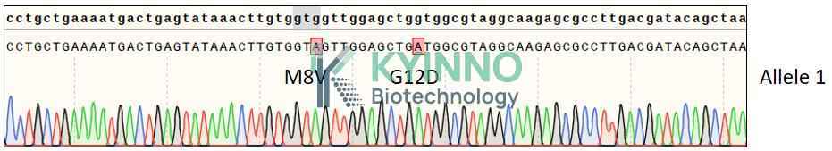
Kirsten rat sarcoma virus (KRAS) is the most frequently mutated oncogene in human cancers, with codon 12 mutations accounting for the majority of activating alterations. Among these, the glycine‑to‑aspartic acid substitution at position 12 (p.G12D) is the most common KRAS mutant allele in pancreatic ductal adenocarcinoma (PDAC, ~37–40%) and colorectal cancer (CRC, ~13–15%), and is also recurrent in non‑small cell lung cancer (NSCLC, ~4%) as well as ampullary and biliary tract cancers. The G12D mutation resides in the phosphate‑binding loop of the GTPase domain and impairs intrinsic GTP hydrolysis, leading to persistent activation of downstream RAF‑MEK‑ERK (MAPK) and PI3K‑AKT signaling, which drives malignant proliferation, metabolic reprogramming, and immune evasion.
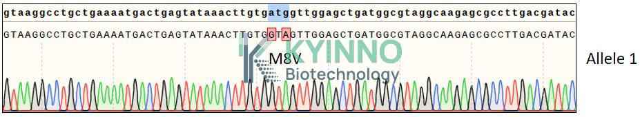
mKRAS-M8V is a missense mutation in KRAS that substitutes methionine (M) with valine (V) at amino acid position 8. This evolutionarily conserved residue is located upstream of the membrane-binding region. Structural predictions indicate that the M8V mutation may mildly perturb the conformation of the KRAS protein, and computational tools such as AlphaMissense classify this variant as likely pathogenic (mean score 0.85). Experimental studies have shown that the M8V mutation moderately enhances the basal GTPase activity of KRAS and promotes the proliferative capacity of colon cancer cells, suggesting that it may act as a weak activating mutation in tumorigenesis. Although this mutation is rare in clinical cohorts, elucidating its functional characteristics contributes to the functional classification of the KRAS mutation spectrum and provides a theoretical basis for developing targeted therapeutic strategies against rare KRAS mutant subtypes.

Specifications

Catalog NumberKC-6142
Cell Line NameCT26-mKRAS-V8-G12D-KI cell line
Clone Number5A1
Host Cell LineCT26
DescriptionStable CT26 clone expressing endogenous KRAS gene bearing G12D mutations
QuantityTwo vials of frozen cells (≥2-106/vial)
StabilityStable in culture over a minimum of 10 passages
ApplicationDrug screening and biological assays
Freezing Medium70% RPMI1640 + 20% FBS + 10% DMSO
Propagation MediumRPMI1640 + 10% FBS
Selection MarkerNA
MorphologyFibroblastoid cells growing as a monolayer
SubcultureSplit the saturated culture at a ratio of 1:4-1:5 every 3-4 days; seed out at about 1-3 x 10⁵ cells/mL
Incubation37 °C with 5% CO2
StorageLiquid nitrogen immediately upon receiving
Doubling TimeApproximately 28 hours
Mycoplasma StatusNegative

Cell Line Generation

CT26-mKRAS-V8-G12D-KI cell line was generated using the CRISPR method.

Characterization

Figure 1: Characterization of CT26-mKRAS-V8-G12D-KI cell line stable clone using PCR sequencing.

Figure 2:Characterization of CT26-mKRAS-V8-G12D-KI cell line stable clone using RT-PCR sequencing.

Figure 3: Characterization of Dose-response curves and IC50 values for CT26 and CT26-mKRAS-V8-G12D-KI cells treated with RMC-6236, BI-2865, AMG510, MRTX-849 and MRTX-1133 over 5 days

Cell Resuscitation

  1. Prewarm culture medium (RPMI1640+10% FBS)in a 37°C water bath.
  2. Thaw the frozen vial in a 37°C water bath for 1-2 minutes.
  3. Transfer the vial into biosafety cabinet, and wipe the surface with 70% ethanol.
  4. Unscrew the top of the vial and transfer the cell suspension gently into a sterile centrifuge tube containing 9.0mL complete culture medium.
  5. Spin at ~ 125 × g for 5-7 minutes at room temperature, and discard the supernatant without disturbing the pellet.
  6. Resuspend cell pellet with the appropriate volume of complete medium and transfer the cell suspension into a T25 culture flask.
  7. Incubate the flask at 37°C, 5% CO2 incubator.
  8. Split saturated culture 1:4-1:5 every 3-4 days; seed out at about 1-3 × 10⁵ cells/mL.

Cell Freezing

  1. Prepare the freezing medium (70% RPMI1640 + 20% FBS + 10% DMSO) fresh immediately before use.
  2. Keep the freezing medium on ice and label cryovials.
  3. Transfer cells to a sterile, conical centrifuge tube, and count the cells.
  4. Centrifuge the cells at 250×g for 5 minutes at room temperature and carefully aspirate off the medium.
  5. Resuspend the cells at a density of at least 3×10⁶ cells/mL in chilled freezing medium.
  6. Aliquot 1 mL of the cell suspension into each cryovial.
  7. Freeze cells in the CoolCell freezing container overnight in a -80°C freezer.
  8. Transfer vials to liquid nitrogen for long-term storage.

References

  1. Shi K, Li A. Targeting KRAS G12D: Advances in Inhibitor Design. Thorac Cancer. 2025 Dec;16(24):e70203. doi: 10.1111/1759-7714.70203. PMID: 41423807; PMCID: PMC12719395.
  2. Wu X, Hayashi H, Tanizaki T, Sun Y, Iwatsuki M. Emerging KRAS G12D inhibitor in the treatment of digestive system tumors: opportunities and challenges. Transl Gastroenterol Hepatol. 2026 Mar 20;11:49. doi: 10.21037/tgh-25-124. PMID: 41969551; PMCID: PMC13066353.

Use License Agreement

Research Use Only.
Not for use in diagnostic procedures or therapeutic applications.
Redistribution of the cell line or its derivatives is prohibited without prior written permission from Kyinno Biotechnology.
请在浏览器中启用JavaScript来完成此表单。