KC-3339

293T-cyno-CLEC4C-FCER1G Cell Line

×
Please enable JavaScript in your browser to complete this form.
26965
Home » 293T-cyno-CLEC4C-FCER1G Cell Line

Background of 293T-cyno-CLEC4C-FCER1G Cell Line

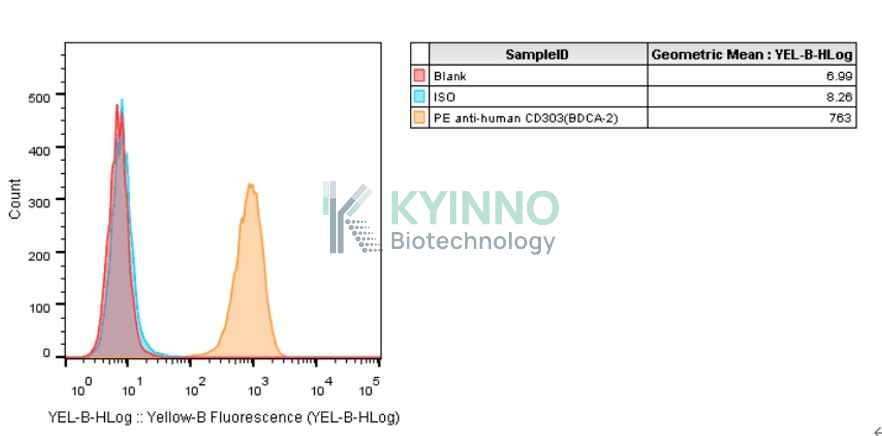
CLEC4C (also known as BDCA-2 or CD303) is the fourth member of the C-type lectin domain family 4 member C. CLEC4C was mainly expressed on the surface of plasmacytoid dendritic cells (pDC). As one of the markers of plasma dendritic cells, CLEC4C plays an important role in the immune system, especially in autoimmune diseases such as systemic lupus erythematosus (SLE). The study suggests that CLEC4C may effectively inhibit the induction of interferon α/β production in plasmacytoid DCS through a mechanism dependent on calcium mobilization and protein tyrosine. Because plasmacytoid dendritic cells produce interferon-α /β, it is considered to be a major pathophysiological factor in systemic lupus erythematosus. Therefore, CLEC4C/BDCA-2 becomes a potential target for blocking the production of interferon-α /β in SLE.
Fc epsilon receptor Ig (FCER1G) is a gene located on chromosome 1 at position 1q23.3 that was first described in 1990. It encodes the γ chain of the Fc receptor, which is the third subunit of the high-affinity immunoglobulin E (IgE) receptor (Fcε RI). The Fc receptor is a protein found on the surface of many different cells: neutrophils, basophils, eosinophils, platelets, macrophages, B lymphocytes, NK cells, mast cells, and dendritic cells. FCER1G expression has been studied and described in various diseases, including squamous cell carcinoma, eczema, meningioma, leukemia, glioma, kidney disease, and even in acute myocardial infarction. Currently, there are four drugs targeting CLEC4C/BDCA-2 in the world (Litifilimab; DB-2304; CBS-004; CDX-1402), and BIIB-059 is the only BDCA-2 mab in the world entering clinical trials.

Specifications

Catalog NumberKC-3339
Cell Line Name293T-cyno-CLEC4C-FCER1G Cell Line
Host Cell Line293T
DescriptionStable HEK293T clone expressing exogenous cyno CLEC4C-FCER1G gene
QuantityTwo vials of frozen cells (≥2-106/vial)
StabilityStable in culture over a minimum of 10 passages
ApplicationDrug screening and biological assays
Freezing Medium70% DMEM + 20% FBS + 10% DMSO
Propagation MediumDMEM + 10% FBS + 1μg/mL Puromycin
Selection MarkerPuromycin
MorphologyEpithelial
SubcultureSplit saturated culture 1:4-1:8 every 2-3 days; seed out at about 1-3 × 10⁵ cells/mL
Incubation37 °C with 5% CO2
StorageLiquid nitrogen immediately upon receiving
Doubling TimeApproximately 30 hours
Mycoplasma StatusNegative
In Vivo ValidationNA

Cell Line Generation

293T cyno-CLEC4C-FCER1G cell line was generated using a lentiviral vector expressing the cyno CLEC4C and FCER1G sequence.

Characterization

Figure 1: Characterization of cyno CLEC4C overexpression in the 293T cyno-CLEC4C-FCER1G stable clone using FACS.

Figure 2: Characterization of endogenous CLEC4C expression in 293T cells using FACS.

Figure 3: Characterization of cyno CLEC4C and FCER1G in the 293T cyno-CLEC4C-FCER1G stable clone using PCR sequencing.

Cell Resuscitation

1. Prewarm culture medium (DMEM supplemented with 10% FBS and 1μg/mL puromycin)in a 37°C water bath.
2. Thaw the frozen vial in a 37°C water bath for 1-2 minutes.
3. Transfer the vial into biosafety cabinet, and wipe the surface with 70% ethanol.
4. Unscrew the top of the vial and transfer the cell suspension gently into a sterile centrifuge tube containing 9.0mL complete culture medium.
5. Spin at ~ 125 × g for 5-7 minutes at room temperature, and discard the supernatant without disturbing the pellet.
6. Resuspend cell pellet with the appropriate volume of complete medium and transfer the cell suspension into a T25 culture flask.
7. Incubate the flask at 37°C, 5% CO2 incubator.
8. Split saturated culture 1:4-1:8 every 2-3 days; seed out at about 1-3 × 10⁵ cells/mL.

Cell Freezing

1. Prepare the freezing medium (70% DMEM + 20% FBS + 10% DMSO) fresh immediately before use.
2. Keep the freezing medium on ice and label cryovials.
3. Transfer cells to a sterile, conical centrifuge tube, and count the cells.
4. Centrifuge the cells at 250×g for 5 minutes at room temperature and carefully aspirate off the medium.
5. Resuspend the cells at a density of at least 3×10⁶ cells/mL in chilled freezing medium.
6. Aliquot 1 mL of the cell suspension into each cryovial.
7. Freeze cells in the CoolCell freezing container overnight in a -80°C freezer.
8. Transfer vials to liquid nitrogen for long-term storage.

References

1. Podgórska D, Cieśla M, Kolarz B. FCER1G Gene Hypomethylation in Patients with Rheumatoid Arthritis. J Clin Med. 2022 Aug 9;11(16):4664. doi: 10.3390/jcm11164664. PMID: 36012903; PMCID: PMC9410058.
2. Reizis B. Regulation of plasmacytoid dendritic cell development. Curr Opin Immunol. 2010 Apr;22(2):206-11. doi: 10.1016/j.coi.2010.01.005. Epub 2010 Feb 9. PMID: 20144853; PMCID: PMC2854232.
3. Toma-Hirano M, Namiki S, Miyatake S, Arai K, Kamogawa-Schifter Y. Type I interferon regulates pDC maturation and Ly49Q expression. Eur J Immunol. 2007 Oct;37(10):2707-14. doi: 10.1002/eji.200737173. PMID: 17823983.
4. Jégouzo, Sabine AF,et al.A novel mechanism for binding of galactose-terminated glycans by the C-type carbohydrate recognition domain in blood dendritic cell antigen 2. Journal of Biological Chemistry 290.27 (2015): 16759-16771.
Please enable JavaScript in your browser to complete this form.