KC-3271

293T-PTH1R Cell Line

×
Please enable JavaScript in your browser to complete this form.
26479
Home » 293T-PTH1R Cell Line

Background of 293T-PTH1R Cell Line

PTH1R, also known as PTHR1, is a protein that belongs to the G-protein coupled receptor 2 family. PTH1R is a receptor for parathyroid hormone and for parathyroid hormone-related peptide. The activity of this receptor is mediated by G proteins which activate adenylyl cyclase, and phosphatidylinositol-calcium second messenger system. Diseases associated with PTH1R include Jansen type metaphyseal chondrodysplasia and Blomstrand type Chondrodysplasia. Among its related pathways are endochondral ossification with skeletal dysplasias and GPCR downstream signaling.

Specifications

Catalog NumberKC-3271
Cell Line Name293T-PTH1R Cell Line
Host Cell Line293T
DescriptionStable 293T cell line expressing exogenous human PTH1R gene
QuantityTwo vials of frozen cells (≥2-106/vial)
StabilityStable in culture over a minimum of 10 passages
ApplicationDrug screening and biological assays
Freezing Medium70% DMEM + 20% FBS + 10% DMSO
Propagation MediumDMEM + 10% FBS + 1µg/mL puromycin
Selection MarkerPuromycin
MorphologyEpithelial
SubcultureSplit saturated culture 1:4-1:5 every 2-3 days; seed out at about 1-2 × 10⁵ cells/ml
Incubation37 °C with 5% CO2
StorageLiquid nitrogen immediately upon receiving
Doubling TimeApproximately 30 hours
Mycoplasma StatusNegative
In Vivo ValidationNA

Cell Line Generation

293T human PTH1R Cell Line was generated using a lentiviral vector expressing the human PTH1R sequence.

Characterization

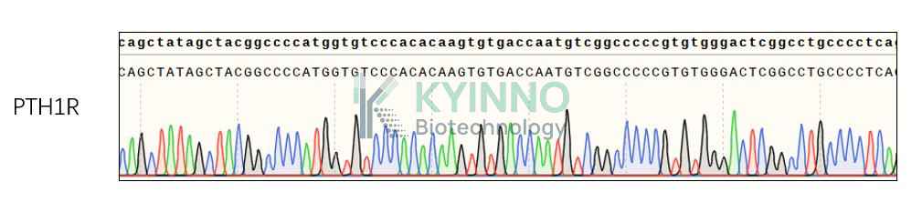
Figure 1: Characterization of human PTH1R overexpression in the 293T human PTH1R stable clone using PCR sequence

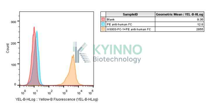
Figure 2: Characterization of human PTH1R overexpression in the 293T human PTH1R stable clone using FACS.

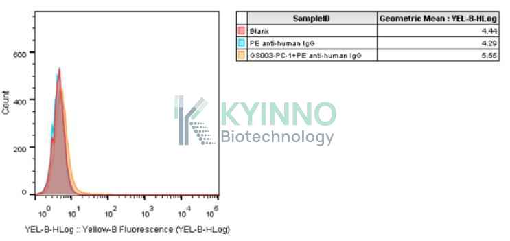
Figure3: Characterization of endogenous human PTH1R expression in 293T using FACS.

Cell Resuscitation

1. Prewarm culture medium (DMEM + 10% FBS + 1µg/mL puromycin)in a 37°C water bath.
2. Thaw the frozen vial in a 37°C water bath for 1-2 minutes.
3. Transfer the vial into biosafety cabinet, and wipe the surface with 70% ethanol.
4. Unscrew the top of the vial and transfer the cell suspension gently into a sterile centrifuge tube containing 9.0mL complete culture medium.
5. Spin at ~ 125 × g for 5-7 minutes at room temperature, and discard the supernatant without disturbing the pellet.
6. Resuspend cell pellet with the appropriate volume of complete medium and transfer the cell suspension into a T25 culture flask.
7. Incubate the flask at 37°C, 5% CO2 incubator.
8. Split saturated culture 1:4-1:5 every 2-3 days; seed out at about 1-2 × 10⁵ cells/mL.

Cell Freezing

1. Prepare the freezing medium (70% DMEM + 20% FBS + 10% DMSO) fresh immediately before use.
2. Keep the freezing medium on ice and label cryovials.
3. Transfer cells to a sterile, conical centrifuge tube, and count the cells.
4. Centrifuge the cells at 250×g for 5 minutes at room temperature and carefully aspirate off the medium.
5. Resuspend the cells at a density of at least 3×10⁶ cells/mL in chilled freezing medium.
6. Aliquot 1 mL of the cell suspension into each cryovial.
7. Freeze cells in the CoolCell freezing container overnight in a -80°C freezer.
8. Transfer vials to liquid nitrogen for long-term storage.

References

1. Structure of the parathyroid hormone receptor C terminus bound to the G-protein dimer Gbeta1gamma2. PMID: 18611381 PMCID: PMC2601695
2. Dimeric arrangement of the parathyroid hormone receptor and a structural mechanism for ligand-induced dissociation. PMID: 20172855 PMCID: PMC2852981
3. Yamaguchi T, Hosomichi K, Narita A, Shirota T, Tomoyasu Y, Maki K, Inoue I (Jul 2011). Exome resequencing combined with linkage analysis identifies novel PTH1R variants in primary failure of tooth eruption in Japanese. Journal of Bone and Mineral Research. 26 (7): 1655–61.
Please enable JavaScript in your browser to complete this form.