KC-0203

293T-Wnt-Luciferase-Reporter-Cell-Line

×
Please enable JavaScript in your browser to complete this form.
20005
Home » 293T-Wnt-Luciferase-Reporter-Cell-Line

Background of 293T-Wnt-Luciferase-Reporter-Cell-Line

NFκB signaling plays a critical role in developmental process, including cell proliferation, differentiation and tissue patterning, and can be aberrantly activated in cancer and diabetes. When Wnt pathway is activated, the β-catenin protein complex translocate from cytoplasm to nucleus, regulates a wide spectrum of gene expression after binds to its response element on the promoter region of downstream gene.

Specifications

Catalog NumberKC-0203
Cell Line Name293T-Wnt-Luciferase-Reporter-Cell-Line
Host Cell Line293T
DescriptionHEK293T cell line stable expressing exogenous firefly luciferase gene under the
control of TCF-responsive element
QuantityTwo vials of frozen cells (≥2-106/vial)
StabilityStable in culture over a minimum of 10 passages
ApplicationDrug screening and biological assays
Freezing Medium70% DMEM + 20% FBS + 10% DMSO
Propagation MediumDMEM + 10% FBS
Selection MarkerNA
MorphologyEpithelial
SubcultureSplit saturated culture 1:4-1:5 every 2-3 days; seed out at about 1-3 × 10⁵ cells/mL
Incubation37 °C with 5% CO2
StorageLiquid nitrogen immediately upon receiving
Doubling TimeApproximately 30 hours
Mycoplasma StatusNegative
In Vivo ValidationNA

Cell Line Generation

293T Wnt luciferase cell line was generated using plasmid transfection method.

Characterization

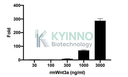
Figure: 293T Wnt luciferase cell line was seed into the 96-well plate, and treated with rmWnt3a for 16 hours, then readout with Bright-Glo system

Cell model for monitoring Wnt pathway.

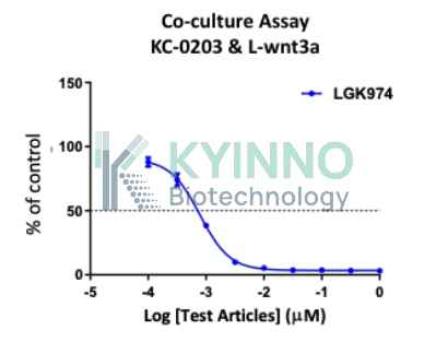
Example: PORCN inhibitor test in co-culture reporter assay of 293-wnt-reporter line and L-wnt3a line

Cell Resuscitation

  1. Prewarm culture medium (DMEM supplemented with 10% FBS)in a 37°C water bath.
  2. Thaw the frozen vial in a 37°C water bath for 1-2 minutes.
  3. Transfer the vial into biosafety cabinet, and wipe the surface with 70% ethanol.
  4. Unscrew the top of the vial and transfer the cell suspension gently into a sterile centrifuge tube containing 9.0mL complete culture medium.
  5. Spin at ~ 125 × g for 5-7 minutes at room temperature, and discard the supernatant without disturbing the pellet.
  6. Resuspend cell pellet with the appropriate volume of complete medium and transfer the cell suspension into a T25 culture flask.
  7. Incubate the flask at 37°C, 5% CO2 incubator.
  8. Split saturated culture 1:4-1:5 every 2-3 days; seed out at about 1-3 × 10⁵ cells/mL.

Cell Freezing

  1. Prepare the freezing medium (70% DMEM + 20% FBS + 10% DMSO) fresh immediately before use.
  2. Keep the freezing medium on ice and label cryovials.
  3. Transfer cells to a sterile, conical centrifuge tube, and count the cells.
  4. Centrifuge the cells at 250×g for 5 minutes at room temperature and carefully aspirate off the medium.
  5. Resuspend the cells at a density of at least 3×10⁶ cells/mL in chilled freezing medium.
  6. Aliquot 1 mL of the cell suspension into each cryovial.
  7. Freeze cells in the CoolCell freezing container overnight in a -80°C freezer.
  8. Transfer vials to liquid nitrogen for long-term storage

References

  1. MacDonald, B. T., Tamai, K. & He, X. Wnt/β-Catenin Signaling: Components, Mechanisms, and Diseases. Dev.
  2. Cell 17, 9–26 (2009).
  3. Nile, A. H. & Hannoush, R. N. Fatty acylation of Wnt proteins. Nat. Chem. Biol. 12, 60–69 (2016).
  4. Cheng, Y. et al. Wnt-C59 arrests stemness and suppresses growth of nasopharyngeal carcinoma in mice by
  5. inhibiting the Wnt pathway in the tumor microenvironment. Oncotarget 6, 14428–14439 (2015).
Please enable JavaScript in your browser to complete this form.