KC-4694

3A9-mouse-CD3E-KO-4C2 Cell Line

×
Please enable JavaScript in your browser to complete this form.
49459
Home » 3A9-mouse-CD3E-KO-4C2 Cell Line

Background of 3A9-mouse-CD3E-KO-4C2 Cell Line

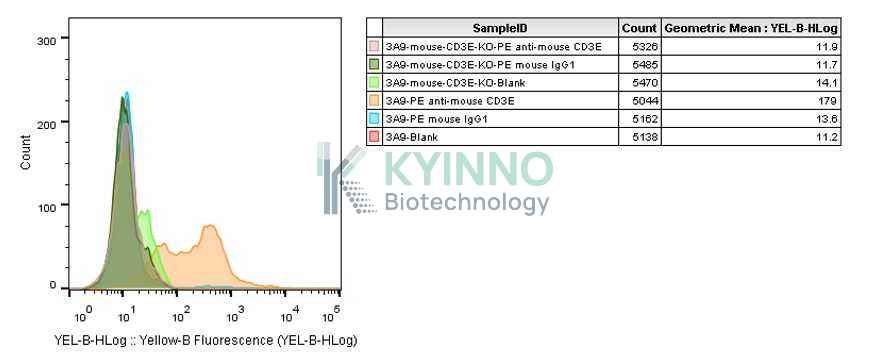
The CD3E gene encodes a component of the T-cell receptor (TCR) complex, which plays a critical role in the activation and function of T cells, an essential part of the adaptive immune system. In mice, the CD3E protein is one of five invariant chains that associate with the variable αβ heterodimer to form the complete TCR complex on the surface of T cells. This complex is crucial for recognizing antigens presented by major histocompatibility complex (MHC) molecules, thereby initiating downstream signaling pathways that lead to T cell activation, proliferation, and differentiation.Mutations or deficiencies in the CD3E gene can result in impaired T cell development and function, leading to immunodeficiency disorders. Studies have shown that CD3E-deficient mice exhibit severe defects in T cell maturation and are highly susceptible to infections. The CD3E protein also interacts with other members of the CD3 family (CD3γ, CD3δ, and CD3ζ), forming a signaling platform that transmits antigen recognition signals into the cell interior.

Specifications

Catalog NumberKC-4694
Cell Line Name3A9-mouse-CD3E-KO-4C2 Cell Line
Host Cell Line3A9
DescriptionStable 3A9 clone with mouse CD3E gene knockout, No.4C2
QuantityTwo vials of frozen cells (≥2-106/vial)
StabilityStable in culture over a minimum of 10 passages
ApplicationDrug screening and biological assays
Freezing MediumRPMI1640+20% FBS+10% DMSO
Propagation MediumRPMI1640+10% FBS
Selection MarkerNA
MorphologyLymphoblast
SubcultureSplit saturated culture 1:5-1:10 every 1-2 days; seed out at about 1-3 × 10⁵ cells/mL
Incubation37 °C with 5% CO2
StorageLiquid nitrogen immediately upon receiving
Doubling TimeApproximately 16 hours
Mycoplasma StatusNegative

Cell Line Generation

3A9-mouse-CD3E-KO-4C2 cell line was generated using the CRISPR method.

Characterization

Figure 1: Characterization of CD3E knockout in 3A9 using PCR sequencing.

Figure 2: Characterization of CD3E knockout in 3A9 using RT-PCR sequencing.

Figure 3: Characterization of CD3E knockout in 3A9 using FACS.

Cell Resuscitation

  1. Prewarm culture medium (RPMI1640 + 10% FBS)in a 37°C water bath.
  2. Thaw the frozen vial in a 37°C water bath for 1-2 minutes.
  3. Transfer the vial into biosafety cabinet, and wipe the surface with 70% ethanol.
  4. Unscrew the top of the vial and transfer the cell suspension gently into a sterile centrifuge tube containing 9.0mL complete culture medium.
  5. Spin at ~ 125 × g for 5-7 minutes at room temperature, and discard the supernatant without disturbing the pellet.
  6. Resuspend cell pellet with the appropriate volume of complete medium and transfer the cell suspension into a T25 culture flask.
  7. Incubate the flask at 37°C, 5% CO2 incubator.
  8. Split saturated culture 1:5-1:10 every 1-2 days; seed out at about 1-3 × 10⁵ cells/mL.

Cell Freezing

  1. Prepare the freezing medium (70% RPMI-1640 + 20% FBS + 10% DMSO) fresh immediately before use.
  2. Keep the freezing medium on ice and label cryovials.
  3. Transfer cells to a sterile, conical centrifuge tube, and count the cells.
  4. Centrifuge the cells at 250×g for 5 minutes at room temperature and carefully aspirate off the medium.
  5. Resuspend the cells at a density of at least 3×10⁶ cells/mL in chilled freezing medium.
  6. Aliquot 1 mL of the cell suspension into each cryovial.
  7. Freeze cells in the CoolCell freezing container overnight in a -80°C freezer.
  8. Transfer vials to liquid nitrogen for long-term storage

References

1.Call ME, Pyrdol J, Wiedmann M, Wucherpfennig KW. The organizing principle in the formation of the T cell receptor-CD3 complex. Cell. 2002 Dec 27;111(7):967-79. doi: 10.1016/s0092-8674(02)01194-7. PMID: 12507424; PMCID: PMC3420808.
2.Davis SJ, van der Merwe PA. The kinetic-segregation model: TCR triggering and beyond. Nat Immunol. 2006 Aug;7(8):803-9. doi: 10.1038/ni1369. PMID: 16855606.
3.Dustin ML, Chan AC. Signaling takes shape in the immune system. Cell. 2000 Oct 13;103(2):283-94. doi: 10.1016/s0092-8674(00)00120-3. PMID: 11057901.
Please enable JavaScript in your browser to complete this form.