KC-3783

Ba/F3-MPL-CALR-del61

Stable Ba/F3 cell line expressing exogenous MPL & CALR-del61 gene

×
请在浏览器中启用JavaScript来完成此表单。
33868
Home » Ba/F3-MPL-CALR-del61

Background of Ba/F3-MPL-CALR-del61

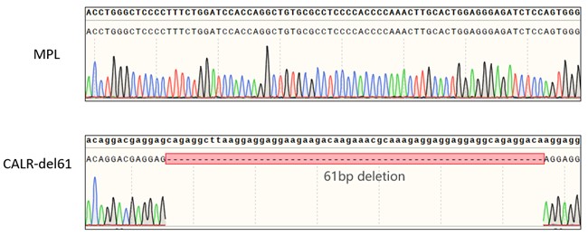
MPL (Thrombopoietin Receptor/CD110) is a cytokine receptor essential for hematopoietic stem cell function, while CALR (Calreticulin) is an endoplasmic reticulum chaperone. Driver mutations in MPN include JAK2, MPL, and CALR exon 9 frameshifts (e.g., del52, ins5, del61). Mutant CALR binds MPL, inducing constitutive JAK-STAT signaling. Approved therapy includes JAK inhibitors (ruxolitinib); novel agents targeting the mutant CALR-MPL complex (e.g., INCA033989) are in clinical trials.

Specifications

Catalog NumberKC-3783
Cell Line NameBa/F3-MPL-CALR-del61
NCBI/UniProt Accession NumberNM_005373, NM_004343.4
Clone Number1#
Host Cell LineMouse Ba/F3 cell line
DescriptionStable Ba/F3 cell line expressing exogenous human MPL and del61 mutant form of CALR gene.
QuantityOne vial of frozen cells (≥2-106/vial)
StabilityStable in culture over a minimum of 10 passages
ApplicationDrug screening and biological assays
Freezing Medium70% RPMI1640+20% FBS+10% DMSO
Propagation MediumRPMI1640+10%FBS
Selection MarkerPuromycin | G418
MorphologyMostly single, round (some polymorph) cells in suspension
SubcultureSplit saturated culture 1:10 every 3 days
Incubation37 °C with 5% CO2
StorageLiquid nitrogen immediately upon receiving
Doubling TimeApproximately 20 hours
Mycoplasma StatusNegative
In Vivo ValidationYes

Cell Line Generation

Ba/F3-MPL-CALR-del61 cell line was generated using a retrovirus expressing the human MPL and del61 mutant form of CALR sequence.

Characterization

Figure: Characterization of the BaF3-MPL-CALR-del61 Cell Line stable clone using PCR sequencing.

Cell Resuscitation

  1. Pre-warm complete culture medium (basal medium and 10% FBS) in a 37°C water bath.
  2. Rapidly thaw the cryovial in a 37°C water bath for 1-2 minutes with gentle agitation.
  3. Transfer the vial to a biosafety cabinet, and disinfect the exterior with 70% ethanol.
  4. Aseptically transfer the cell suspension dropwise into a sterile centrifuge tube containing 9.0 mL of pre-warmed complete medium.
  5. Centrifuge at approximately 125 × g for 5–7 minutes at room temperature, carefully aspirate the supernatant without disturbing the cell pellet.
  6. Gently resuspend the pellet in an appropriate volume of complete medium and transfer the suspension into a T25 flask.
  7. Incubate the flask in a 37°C in a humidified 5% CO2 incubator.
  8. Assess cell viability and morphology after 24 hours. If cells appear healthy, replace the medium with fresh medium supplemented with the appropriate selective antibiotic.
  9. Subculture the cells at a ratio of 1:10 every 3 days upon reaching 80%–90% confluency.

Cell Freezing

  1. Prepare the freezing medium (70% basal medium, 20% FBS and 10% DMSO) freshly before use.
  2. Pre-chill the freezing medium on ice and label the cryovials accordingly.
  3. Transfer the cell suspension to a sterile conical tube and perform a cell count to determine total viability and density.
  4. Centrifuge the cells at 250×g for 5 minutes at room temperature; carefully aspirate the supernatant.
  5. Gently resuspend the cell pellet in chilled freezing medium, ensuring a minimum cell density of 3×10⁶ cells/mL.
  6. Aliquot 1 mL of the cell suspension into each pre-labeled cryovial.
  7. Place the cryovials into a CoolCell® container and store at -80°C overnight for controlled-rate cooling.
  8. Transfer the cryovials to the liquid nitrogen for long-term storage the following day.

References

1. Lim, K-H et al. “Expression of CALR mutants causes mpl-dependent thrombocytosis in zebrafish.” Blood cancer journal vol. 6,10 e481. 7 Oct. 2016, doi:10.1038/bcj.2016.83.
2. Toppaldoddi, Katte Rao et al. “Rare type 1-like and type 2-like calreticulin mutants induce similar myeloproliferative neoplasms as prevalent type 1 and 2 mutants in mice.” Oncogene vol. 38,10 (2019): 1651-1660. doi:10.1038/s41388-018-0538-z.
3. Nieborowska-Skorska, Margaret et al. “Ruxolitinib-induced defects in DNA repair cause sensitivity to PARP inhibitors in myeloproliferative neoplasms.” Blood vol. 130,26 (2017): 2848-2859. doi:10.1182/blood-2017-05-784942.

Use License Agreement

Research Use Only.
Not for use in diagnostic procedures or therapeutic applications.
Redistribution of the cell line or its derivatives is prohibited without prior written permission from Kyinno Biotechnology.
请在浏览器中启用JavaScript来完成此表单。