KC-3914

Ba/F3-MPL-CALR-Cell-Line

Explore the BA F3 MPL CALR Cell Line (KC-3914) at Kyinno. Ideal for studying the interaction of MPL and CALR mutations in cellular processes. Quality tested for robust research outcomes.

×
请在浏览器中启用JavaScript来完成此表单。
35024
Home » Ba/F3-MPL-CALR-Cell-Line

Background of Ba/F3-MPL-CALR-Cell-Line

The MPL gene provides instructions for making the thrombopoietin receptor protein, which promotes the growth and division (proliferation) of cells. This receptor is especially important for the proliferation of certain blood cells called megakaryocytes, which produce platelets, the cells involved in blood clotting. Research suggests that the thrombopoietin receptor may also play a role in the maintenance of hematopoietic stem cells, which are stem cells located within the bone marrow that have the potential to develop into red blood cells, white blood cells, and platelets. The thrombopoietin receptor is turned on (activated) when a protein called thrombopoietin attaches (binds) to it. The activated thrombopoietin receptor stimulates a signaling pathway called the JAK/STAT pathway, which transmits chemical signals from outside the cell to the cell's nucleus and is important for controlling the production of blood cells. Calreticulin, also known as calregulin, CRP55, CaBP3, calsequestrin-like protein, and endoplasmic reticulum resident protein 60 (ERp60), is a protein encoded by the CALR gene. Calreticulin is a multifunctional soluble protein that binds Ca2+ ions (a second messenger in signal transduction), rendering it inactive. The Ca2+ is bound with low affinity, but high capacity, and can be released on a signal (see inositol trisphosphate). Calreticulin is located in storage compartments associated with the endoplasmic reticulum and is considered an ER-resident protein. Ba/F3 cell, a murine interleukin-3 dependent pro-B cell line, is a popular system for exploring both kinases and their inhibitors, because some protein kinases can render the Ba/F3 cells to be depended on the activation of the kinases instead of IL-3 supplement, while their inhibitors can antagonize the kinase-dependent growth effects.

Specifications

Catalog NumberKC-3914
Cell Line NameBa/F3-MPL-CALR-Cell-Line
NCBI/UniProt Accession NumberNM_005373, NM_004343.4
Clone Number1#
Host Cell LineMouse Ba/F3 cell line
DescriptionStable Ba/F3 clone expressing exogenous human MPL and CALR genes
QuantityOne vial of frozen cells (≥2-106/vial)
StabilityStable in culture over a minimum of 10 passages
ApplicationDrug screening and biological assays
Freezing Medium70% RPMI1640+20% FBS+10% DMSO
Propagation MediumRPMI1640+10%FBS+1µg/mL Puromycin+ 8ng/mL mouse-IL3
Selection MarkerPuromycin | G418
MorphologyMostly single, round (some polymorph) cells in suspension
SubcultureSplit saturated culture 1:10 every 3 days
Incubation37 °C with 5% CO2
StorageLiquid nitrogen immediately upon receiving
Doubling TimeApproximately 20 hours
Mycoplasma StatusNegative
In Vivo ValidationNA

Cell Line Generation

Ba/F3 MPL-CALR-WT cell Line was generated using a retrovirus vector expressing human MPL-CALR-WT sequence.

Characterization

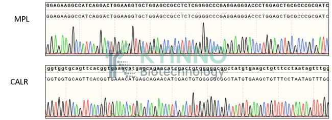
Figure: Characterization of CALR and MPL overexpressing in the Ba/F3 stable clone using PCR sequencing

Cell Resuscitation

  1. Pre-warm complete culture medium (basal medium and 10% FBS) in a 37°C water bath.
  2. Rapidly thaw the cryovial in a 37°C water bath for 1-2 minutes with gentle agitation.
  3. Transfer the vial to a biosafety cabinet, and disinfect the exterior with 70% ethanol.
  4. Aseptically transfer the cell suspension dropwise into a sterile centrifuge tube containing 9.0 mL of pre-warmed complete medium.
  5. Centrifuge at approximately 125 × g for 5–7 minutes at room temperature, carefully aspirate the supernatant without disturbing the cell pellet.
  6. Gently resuspend the pellet in an appropriate volume of complete medium and transfer the suspension into a T25 flask.
  7. Incubate the flask in a 37°C in a humidified 5% CO2 incubator.
  8. Assess cell viability and morphology after 24 hours. If cells appear healthy, replace the medium with fresh medium supplemented with the appropriate selective antibiotic.
  9. Subculture the cells at a ratio of 1:10 every 3 days upon reaching 80%–90% confluency.

Cell Freezing

  1. Prepare the freezing medium (70% basal medium, 20% FBS and 10% DMSO) freshly before use.
  2. Pre-chill the freezing medium on ice and label the cryovials accordingly.
  3. Transfer the cell suspension to a sterile conical tube and perform a cell count to determine total viability and density.
  4. Centrifuge the cells at 250×g for 5 minutes at room temperature; carefully aspirate the supernatant.
  5. Gently resuspend the cell pellet in chilled freezing medium, ensuring a minimum cell density of 3×10⁶ cells/mL.
  6. Aliquot 1 mL of the cell suspension into each pre-labeled cryovial.
  7. Place the cryovials into a CoolCell® container and store at -80°C overnight for controlled-rate cooling.
  8. Transfer the cryovials to the liquid nitrogen for long-term storage the following day.

References

1. Lim, K-H et al. “Expression of CALR mutants causes mpl-dependent thrombocytosis in zebrafish.” Blood cancer journal vol. 6,10 e481. 7 Oct. 2016, doi:10.1038/bcj.2016.83.
2. Toppaldoddi, Katte Rao et al. “Rare type 1-like and type 2-like calreticulin mutants induce similar myeloproliferative neoplasms as prevalent type 1 and 2 mutants in mice.” Oncogene vol. 38,10 (2019): 1651-1660. doi:10.1038/s41388-018-0538-z.
3. Nieborowska-Skorska, Margaret et al. “Ruxolitinib-induced defects in DNA repair cause sensitivity to PARP inhibitors in myeloproliferative neoplasms.” Blood vol. 130,26 (2017): 2848-2859. doi:10.1182/blood-2017-05-784942.

Use License Agreement

Research Use Only.
Not for use in diagnostic procedures or therapeutic applications.
Redistribution of the cell line or its derivatives is prohibited without prior written permission from Kyinno Biotechnology.
请在浏览器中启用JavaScript来完成此表单。