KC-3765

CHOK1-CD66a-short-Cell-Line

×
Please enable JavaScript in your browser to complete this form.
34736
Home » 细胞系 » CHOK1-CD66a-short-Cell-Line

Background of CHOK1-CD66a-short-Cell-Line

CD66a, also known as CEACAM1, is a transmembrane glycoprotein of the immunoglobulin superfamily and a key mediator in various physiological and pathological processes, including tumor diseases. It is expressed in various tissues, including epithelial cells, immune cells, and endothelial cells.

Specifications

Catalog NumberKC-3765
Cell Line NameCHOK1-CD66a-short-Cell-Line
Host Cell LineCHOK1
DescriptionStable CHOK1 clone expressing exogenous CD66a-short gene.
QuantityTwo vials of frozen cells (≥2-106/vial)
StabilityStable in culture over a minimum of 10 passages
ApplicationDrug screening and biological assays
Freezing Medium70% RPMI 1640 + 20% FBS + 10% DMSO
Propagation MediumRPMI 1640 + 10% FBS + 10μg/mL Puromycin
Selection MarkerPuromycin
MorphologyEpithelial
SubcultureSplit saturated culture 1:6-1:8 every 2-3 days; seed out at about 1-3 × 10⁵ cells/mL
Incubation37 °C with 5% CO2
StorageLiquid nitrogen immediately upon receiving
Doubling TimeApproximately 24 hours
Mycoplasma StatusNegative
In Vivo ValidationNA

Cell Line Generation

CHOK1-CD66a-short-cell-line was generated using a lentiviral vector expressing the CD66a-short sequence.

Characterization

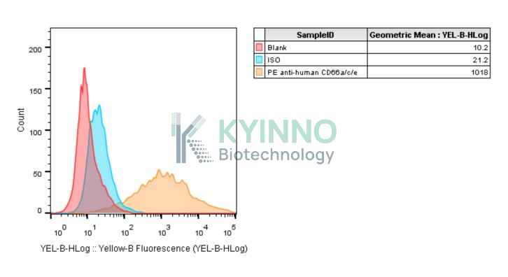
Figure 1: Characterization of CD66a-short overexpression in the CHOK1-CD66a-short stable clone using FACS.

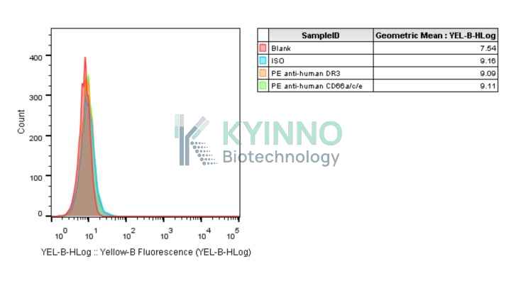
Figure 2: Characterization of CD66a-short overexpression in the CHOK1 stable clone using FACS.

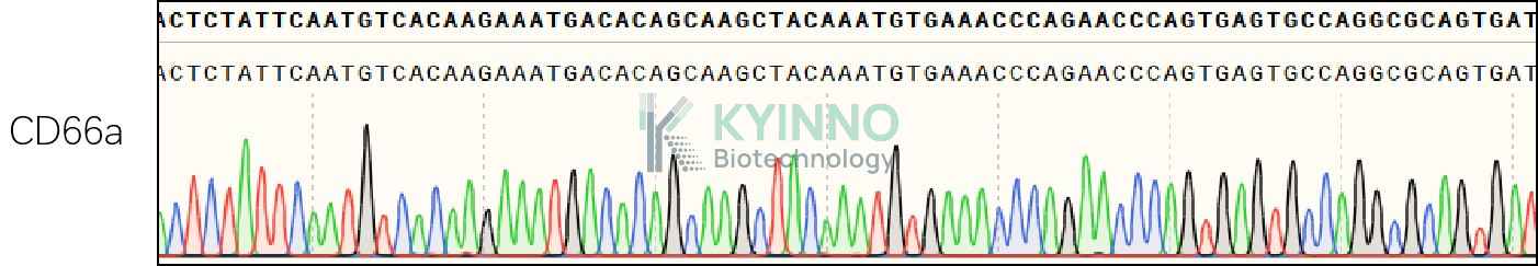
Figure 3: Characterization of CD66a-short in the CHOK1-CD66a-short stable clone using PCR sequencing.

Cell Resuscitation

1. Prewarm culture medium (RPMI 1640 supplemented with 10% FBS and 10μg/mL puromycin)in a 37°C water bath. 2. Thaw the frozen vial in a 37°C water bath for 1-2 minutes. 3. Transfer the vial into biosafety cabinet, and wipe the surface with 70% ethanol. 4. Unscrew the top of the vial and transfer the cell suspension gently into a sterile centrifuge tube containing 9.0mL complete culture medium. 5. Spin at ~ 125 × g for 5-7 minutes at room temperature, and discard the supernatant without disturbing the pellet. 6. Resuspend cell pellet with the appropriate volume of complete medium and transfer the cell suspension into a T25 culture flask. 7. Incubate the flask at 37°C, 5% CO2 incubator. 8. Split saturated culture 1:6-1:8 every 2-3 days; seed out at about 1-3 × 10⁵ cells/mL.

Cell Freezing

1. Prepare the freezing medium (70% RPMI 1640 + 20% FBS + 10% DMSO) fresh immediately before use. 2. Keep the freezing medium on ice and label cryovials. 3. Transfer cells to a sterile, conical centrifuge tube, and count the cells. 4. Centrifuge the cells at 250×g for 5 minutes at room temperature and carefully aspirate off the medium. 5. Resuspend the cells at a density of at least 3×10⁶ cells/mL in chilled freezing medium. 6. Aliquot 1 mL of the cell suspension into each cryovial. 7. Freeze cells in the CoolCell freezing container overnight in a -80°C freezer. 8. Transfer vials to liquid nitrogen for long-term storage.

References

1.Götz L, Rueckschloss U, Najjar SM, Ergün S, Kleefeldt F. Carcinoembryonic antigen-related cell adhesion molecule 1 in cancer: Blessing or curse? Eur J Clin Invest. 2024 Dec;54 Suppl 2(Suppl 2):e14337. doi: 10.1111/eci.14337. Epub 2024 Oct 25. PMID: 39451132; PMCID: PMC11646294. 2. Götz L, Rueckschloss U, Ergün S, Kleefeldt F. CEACAM1 in vascular homeostasis and inflammation. Eur J Clin Invest. 2024 Dec;54 Suppl 2(Suppl 2):e14345. doi: 10.1111/eci.14345. PMID: 39674877; PMCID: PMC11646292.
Please enable JavaScript in your browser to complete this form.