This gene encodes a classical cadherin of the cadherin superfamily. Alternative splicing results in multiple transcript variants, at least one of which encodes a preproprotein that is proteolytically processed to generate the mature glycoprotein. This calcium-dependent cell-cell adhesion protein is comprised of five extracellular cadherin repeats, a transmembrane region and a highly conserved cytoplasmic tail. This gene is located in a gene cluster in a region on the long arm of chromosome 16 that is involved in loss of heterozygosity events in breast and prostate cancer. In addition, aberrant expression of this protein is observed in cervical adenocarcinomas. Mutations in this gene are associated with hypotrichosis with juvenile macular dystrophy and ectodermal dysplasia, ectrodactyly, and macular dystrophy syndrome (EEMS).
Specifications
Catalog Number
KC-3746
Cell Line Name
CHOK1-cyno-CDH3 Cell Line
Host Cell Line
CHOK1
Description
Stable CHOK1 cell line expressing exogenous cyno-CDH3 gene
Quantity
One vial of frozen cells (≥2-106/vial)
Stability
Stable in culture over a minimum of 10 passages
Application
Drug screening and biological assays
Freezing Medium
70% RPMI1640 + 20% FBS + 10% DMSO
Propagation Medium
RPMI1640 + 10% FBS + 10μg/ml Puromycin
Selection Marker
Puromycin
Morphology
Epithelial
Subculture
Split saturated culture 1:4-1:8 every 2-3 days; seed out at about 1-3 × 10⁵ cells/mL
Incubation
37 °C with 5% CO2
Storage
Liquid nitrogen immediately upon receiving
Doubling Time
Approximately 30 hours
Mycoplasma Status
Negative
In Vivo Validation
NA
Cell Line Generation
CHOK1-cyno-CDH3 cell line was generated using lentivirus expressing cyno-CDH3 sequence.
Characterization
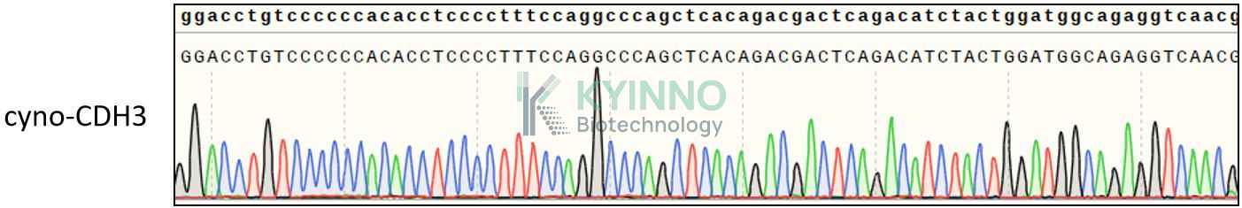
Figure 1. Characterization of cyno-CDH3 over-expression in the CHOK1-cyno-CDH3 stable clone using PCR.
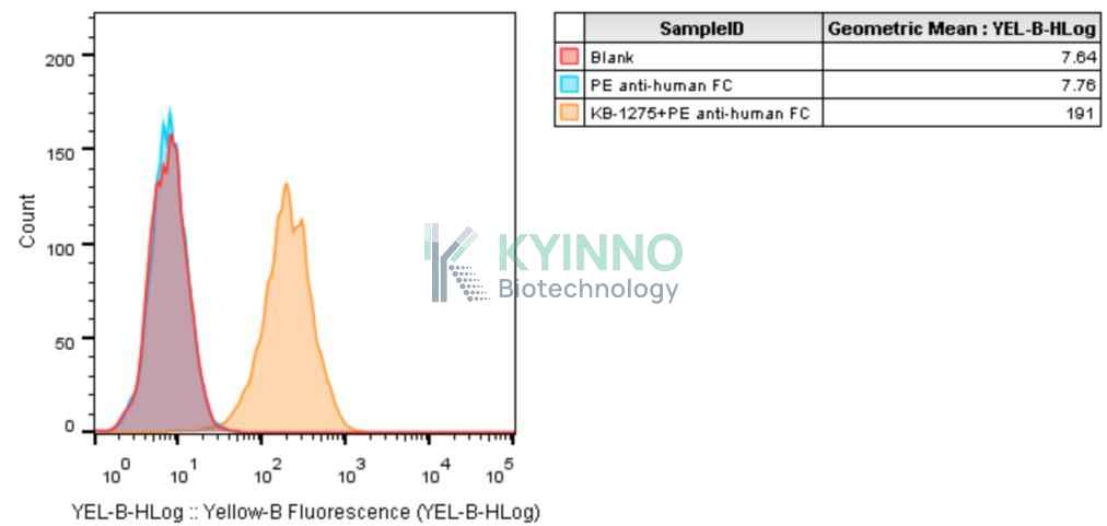
Figure 2. Characterization of cyno-CDH3 over-expression in the CHOK1-cyno-CDH3 stable clone using FACS.
Cell Resuscitation
1. Prewarm culture medium (RPMI1640 supplemented with 10% FBS and 10μg/mL Puromycin)in a 37°C water bath.
2. Thaw the frozen vial in a 37°C water bath for 1-2 minutes.
3. Transfer the vial into biosafety cabinet, and wipe the surface with 70% ethanol.
4. Unscrew the top of the vial and transfer the cell suspension gently into a sterile centrifuge tube containing 9.0mL complete culture medium.
5. Spin at ~ 125 × g for 5-7 minutes at room temperature, and discard the supernatant without disturbing the pellet.
6. Resuspend cell pellet with the appropriate volume of complete medium and transfer the cell suspension into a T25 culture flask.
7. Incubate the flask at 37°C, 5% CO2 incubator.
8. Split saturated culture 1:4-1:8 every 2-3 days; seed out at about 1-3 × 10⁵ cells/mL.
Cell Freezing
1. Prepare the freezing medium (70% RPMI1640 + 20% FBS + 10% DMSO) fresh immediately before use.
2. Keep the freezing medium on ice and label cryovials.
3. Transfer cells to a sterile, conical centrifuge tube, and count the cells.
4. Centrifuge the cells at 250×g for 5 minutes at room temperature and carefully aspirate off the medium.
5. Resuspend the cells at a density of at least 3×10⁶ cells/mL in chilled freezing medium.
6. Aliquot 1 mL of the cell suspension into each cryovial.
7. Freeze cells in the CoolCell freezing container overnight in a -80°C freezer.
8. Transfer vials to liquid nitrogen for long-term storage.
References
1.Sousa B, Ribeiro AS, Nobre AR, Lopes N, Martins D, Pinheiro C, Vieira AF, Albergaria A, Gerhard R, Schmitt F, Baltazar F, Paredes J. The basal epithelial marker P-cadherin associates with breast cancer cell populations harboring a glycolytic and acid-resistant phenotype. BMC Cancer. 2014 Oct 1;14:734. doi: 10.1186/1471-2407-14-734. PMID: 25269858; PMCID: PMC4190447.
Use License Agreement
Research Use Only.
Not for use in diagnostic procedures or therapeutic applications.
Redistribution of the cell line or its derivatives is prohibited without prior written permission from Kyinno Biotechnology.