KC-2958

CHOK1-IL6ST-Cell-Line

×
Please enable JavaScript in your browser to complete this form.
34022
Home » CHOK1-IL6ST-Cell-Line

Background of CHOK1-IL6ST-Cell-Line

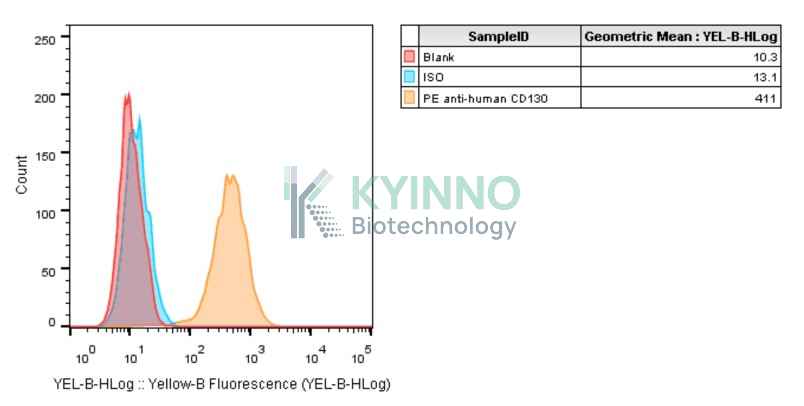
IL6ST, encoding the gp130 protein, serves as a critical shared signaling receptor for the IL-6 cytokine family. By forming functional complexes with various specific receptors, it activates the JAK-STAT pathway to regulate immune responses, hematopoiesis, and cell survival. While essential for homeostasis, its dysregulation is a hallmark of chronic inflammation and solid tumors like CRC and PDAC. Currently, targeting the gp130-mediated trans-signaling pathway with fusion proteins like Olamkicept represents a promising therapeutic strategy for autoimmune diseases and oncology.

Specifications

Catalog NumberKC-2958
Cell Line NameCHOK1-IL6ST-Cell-Line
NCBI/UniProt Accession NumberNM_002184.4
Clone Number2#
Host Cell LineCHOK1
DescriptionStable CHOK1 cell line expressing exogenous human IL6ST gene
QuantityTwo vials of frozen cells (≥2-106/vial)
StabilityStable in culture over a minimum of 10 passages
ApplicationDrug screening and biological assays
Freezing Medium70% RPMI1640+20% FBS+10% DMSO
Propagation MediumRPMI1640+10%FBS+750µg/mL Hygromycin B
Selection MarkerHygromycin B
MorphologyEpithelial-like
SubcultureSplit saturated culture 1:4-1:8 every 2-3 days; seed out at about 1-2 × 10⁵ cells/mL
Incubation37 °C with 5% CO2
StorageLiquid nitrogen immediately upon receiving
Doubling TimeApproximately 30 hours
Mycoplasma StatusNegative
In Vivo ValidationNA

Cell Line Generation

CHOK1-IL6ST Cell Line was generated using a lentiviral vector expressing the human IL6ST sequence.

Characterization

Figure 1: Characterization of IL6ST overexpression in the CHOK1-IL6ST stable clone using FACS.

Cell Resuscitation

1. Prewarm culture medium (RPMI1640 supplemented with 10% FBS and 750μg/mL hygromycin B)in a 37°C water bath.
2. Thaw the frozen vial in a 37°C water bath for 1-2 minutes.
3. Transfer the vial into biosafety cabinet, and wipe the surface with 70% ethanol.
4. Unscrew the top of the vial and transfer the cell suspension gently into a sterile centrifuge tube containing 9.0mL complete culture medium.
5. Spin at ~ 125 × g for 5-7 minutes at room temperature, and discard the supernatant without disturbing the pellet.
6. Resuspend cell pellet with the appropriate volume of complete medium and transfer the cell suspension into a T25 culture flask.
7. Incubate the flask at 37°C, 5% CO2 incubator.
8. Split saturated culture 1:4-1:8 every 2-3 days; seed out at about 1-3 × 10⁵ cells/mL.

Cell Freezing

1. Prepare the freezing medium (70% RPMI1640 + 20% FBS + 10% DMSO) fresh immediately before use.
2. Keep the freezing medium on ice and label cryovials.
3. Transfer cells to a sterile, conical centrifuge tube, and count the cells.
4. Centrifuge the cells at 250×g for 5 minutes at room temperature and carefully aspirate off the medium.
5. Resuspend the cells at a density of at least 3×10⁶ cells/mL in chilled freezing medium.
6. Aliquot 1 mL of the cell suspension into each cryovial.
7. Freeze cells in the CoolCell freezing container overnight in a -80°C freezer.
8. Transfer vials to liquid nitrogen for long-term storage.

References

1. Martínez-Pérez, Carlos et al. “The Signal Transducer IL6ST (gp130) as a Predictive and Prognostic Biomarker in Breast Cancer.” Journal of personalized medicine vol. 11,7 618. 29 Jun. 2021, doi:10.3390/jpm11070618
2. Minegishi, Yoshiyuki. “Hyper-IgE syndrome, 2021 update.” Allergology international : official journal of the Japanese Society of Allergology vol. 70,4 (2021): 407-414. doi:10.1016/j.alit.2021.07.007
Please enable JavaScript in your browser to complete this form.