Enables adipokinetic hormone binding activity and adipokinetic hormone receptor activity. Involved in several processes, including negative regulation of NF-kappaB transcription factor activity; positive regulation of macrophage chemotaxis; and regulation of calcium-mediated signaling. Located in plasma membrane.
Specifications
Catalog Number
KC-5976
Cell Line Name
CHOK1-mouse-CMKLR1 Cell Line
NCBI/UniProt Accession Number
NM_008153.3
Clone Number
5#
Host Cell Line
CHOK1
Description
Stable CHOK1 cell line expressing exogenous mouse CMKLR1 gene
Quantity
One vial of frozen cells (≥2-106/vial)
Stability
Stable in culture over a minimum of 10 passages
Application
Drug screening and biological assays
Freezing Medium
70% basal medium+20% FBS+10% DMSO
Propagation Medium
RPMI 1640+10% FBS +10μg/mL Puromycin
Selection Marker
Puromycin
Morphology
Epithelial-like
Subculture
Split saturated culture 1:4-1:8 every 2-3 days
Incubation
37 °C with 5% CO2
Storage
Liquid nitrogen immediately upon receiving
Doubling Time
Approximately 30 hours
Mycoplasma Status
Negative
In Vivo Validation
NA
Cell Line Generation
CHOK1-mouse-CMKLR1 cell line was generated using a lentiviral vector expressing the mouse CMKLR1 sequence.
Characterization
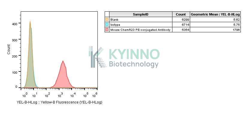
Figure 1: Characterization of mouse CMKLR1 overexpression in the CHOK1-mouse-CMKLR1 stable clone using FACS.
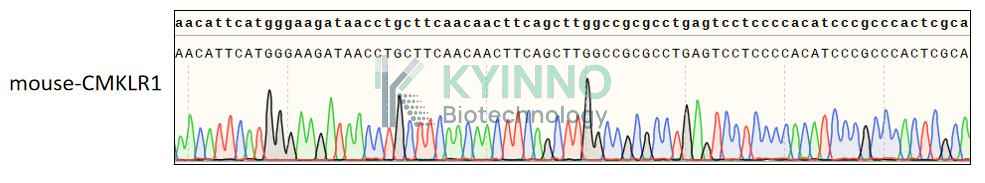
Figure 2: Characterization of mouse CMKLR1 in the CHOK1-mouse-CMKLR1 stable clone using PCR sequencing.
Cell Resuscitation
Pre-warm complete culture medium (basal medium and 10% FBS) in a 37°C water bath.
Rapidly thaw the cryovial in a 37°C water bath for 1-2 minutes with gentle agitation.
Transfer the vial to a biosafety cabinet, and disinfect the exterior with 70% ethanol.
Aseptically transfer the cell suspension dropwise into a sterile centrifuge tube containing 9.0 mL of pre-warmed complete medium.
Centrifuge at approximately 125 × g for 5–7 minutes at room temperature, carefully aspirate the supernatant without disturbing the cell pellet.
Gently resuspend the pellet in an appropriate volume of complete medium and transfer the suspension into a T25 flask.
Incubate the flask in a 37°C in a humidified 5% CO2 incubator.
Assess cell viability and morphology after 24 hours. If cells appear healthy, replace the medium with fresh medium supplemented with the appropriate selective antibiotic.
Subculture the cells at a ratio of 1:4-1:8 every 2-3 days upon reaching 80%–90% confluency.
Cell Freezing
Prepare the freezing medium (70% basal medium, 20% FBS and 10% DMSO) freshly before use.
Pre-chill the freezing medium on ice and label the cryovials accordingly.
Transfer the cell suspension to a sterile conical tube and perform a cell count to determine total viability and density.
Centrifuge the cells at 250×g for 5 minutes at room temperature; carefully aspirate the supernatant.
Gently resuspend the cell pellet in chilled freezing medium, ensuring a minimum cell density of 3×10⁶ cells/mL.
Aliquot 1 mL of the cell suspension into each pre-labeled cryovial.
Place the cryovials into a CoolCell® container and store at -80°C overnight for controlled-rate cooling.
Transfer the cryovials to the liquid nitrogen for long-term storage the following day.
References
1.Li XM, Ji H, Li CJ, Wang PH, Yu P, Yu DM. Chemerin expression in Chinese pregnant women with and without gestational diabetes mellitus. Ann Endocrinol (Paris). 2015 Feb;76(1):19-24.
Use License Agreement
Research Use Only.
Not for use in diagnostic procedures or therapeutic applications.
Redistribution of the cell line or its derivatives is prohibited without prior written permission from Kyinno Biotechnology.