KC-1506

Jurkat-NFAT- Luc2-CD16a-F158 Cell Line

×
Please enable JavaScript in your browser to complete this form.
53442
Home » Jurkat-NFAT- Luc2-CD16a-F158 Cell Line

Background of Jurkat-NFAT- Luc2-CD16a-F158 Cell Line

"CD16, also named FC gamma RIII, is a low or intermediate affinity FC receptor, and has been identified as two receptors including FcγRIIIa (CD16a) and FcγRIIIb (CD16b). It is involved in phagocytosis, secretion of enzymes and inflammatory mediators, antibody-dependent cytotoxicity and clearance of immune complexes. NFAT (nuclear factor of activator T cells) proteins belongs to a family of transcription factors that are very important in T cells activation, differentiation and tolerance. NFAT pathway is activated by Ca2+ and the Ca2+/calmodulin- dependent serine phosphatase calcineurin after the engagement of T cell receptor (TCR) or FC receptor."

Specifications

Catalog NumberKC-1506
Cell Line NameJurkat-NFAT- Luc2-CD16a-F158 Cell Line
Host Cell LineJurkat-NFAT-Luc2
DescriptionStable Jurkat cell line expressing exogenous luciferase under the control of NFAT responsive element and CD16a-F158 fusion sequence.
QuantityTwo vials of frozen cells (≥2-106/vial)
StabilityStable in culture over a minimum of 10 passages
ApplicationDrug screening and biological assays
Freezing Medium70% RPMI1640 + 20% FBS + 10% DMSO
Propagation MediumRPMI1640 + 10% FBS + 300μg/mL Hygromycin B + 0.75μg/mL Puromycin
Selection MarkerHygromycin B and Puromycin
MorphologyLymphoblast
SubcultureSplit saturated culture 1:4-1:5 every 2-3 days; seed out at about 1 × 10⁵ cells/mL
Incubation37 °C with 5% CO2
StorageLiquid nitrogen immediately upon receiving
Doubling TimeApproximately 26 hours
Mycoplasma StatusNegative

Cell Line Generation

Jurkat-NFAT-Luc2-CD16a-F158 Cell Line was generated using a lentiviral vector expressing the human CD16a-F158 sequence.

Characterization

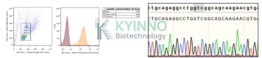
Figure1: Characterization of CD16a-F158 overexpression in Jurkat-NFAT-Luc2-CD16a-F158 stable clones using FACS.

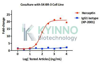
Figure2: Jurkat-NFAT-Luc2-CD16a-F158 and SK-BR-3 cells were seeded into 96-well plates, treated with Herceptin(Cat# KB-1187, Kyinno) in different concentrations for 16 hours, and then read out using Bright-Glo Detection System.

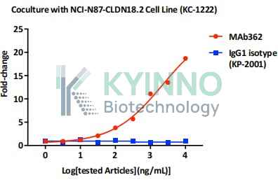
Figure3: Jurkat-NFAT-Luc2-CD16a-F158 and NCI-N87-CLDN18.2 cells were seeded into 96-well plates, treated with Mab362 in different concentrations for 16 hours, and then read out using Bright-Glo Detection System.

Cell Resuscitation

  1. Prewarm culture medium (RPMI 1640 supplemented with 10% FBS, 300µg/mL Hygromycin B and 0.75μg/mL Puromycin) in a 37°C water bath.
  2. Thaw the frozen vial in a 37°C water bath for 1-2 minutes.
  3. Transfer the vial into biosafety cabinet, and wipe the surface with 70% ethanol.
  4. Unscrew the top of the vial and transfer the cell suspension gently into a sterile centrifuge tube containing 9.0mL complete culture medium.
  5. Spin at ~ 125 × g for 5-7 minutes at room temperature, and discard the supernatant without disturbing the pellet.
  6. Resuspend cell pellet with the appropriate volume of complete medium and transfer the cell suspension into a T25 culture flask.
  7. Incubate the flask at 37°C, 5% CO2 incubator.
  8. Split saturated culture 1:4-1:5 every 2-3 days; seed out at about 1 × 10⁵ cells/mL.

Cell Freezing

  1. Prepare the freezing medium (70% RPMI 1640 + 20% FBS + 10% DMSO) fresh immediately before use.
  2. Keep the freezing medium on ice and label cryovials.
  3. Transfer cells to a sterile, conical centrifuge tube, and count the cells.
  4. Centrifuge the cells at 250×g for 5 minutes at room temperature and carefully aspirate off the medium.
  5. Resuspend the cells at a density of at least 3×10⁶ cells/mL in chilled freezing medium.
  6. Aliquot 1 mL of the cell suspension into each cryovial.
  7. Freeze cells in the CoolCell freezing container overnight in a -80°C freezer.
  8. Transfer vials to liquid nitrogen for long-term storage.

References

1. Tada, Minoru, Akiko Ishii-Watabe, Takuo Suzuki, and Nana Kawasaki. 2014. “Development of a Cell-Based Assay Measuring the Activation of FcγRIIa for the Characterization of Therapeutic Monoclonal Antibodies.” Edited by Paul Zhou. PLoS ONE 9 (4): e95787–89. doi:10.1371/journal.pone.0095787.
2. Koene, H R, M Kleijer, J Algra, D Roos, A E von dem Borne, and M de Haas. 1997. “Fc gammaRIIIa-158V/F Polymorphism Influences the Binding of IgG by Natural Killer Cell Fc gammaRIIIa, Independently of the Fc gammaRIIIa-48L/R/H Phenotype..” Blood 90 (3): 1109–14.
Please enable JavaScript in your browser to complete this form.