KC-1507

Jurkat-NFAT- Luc2-CD16a-V158 Cell Line

×
Please enable JavaScript in your browser to complete this form.
22275
Home » Jurkat-NFAT- Luc2-CD16a-V158 Cell Line

Background of Jurkat-NFAT- Luc2-CD16a-V158 Cell Line

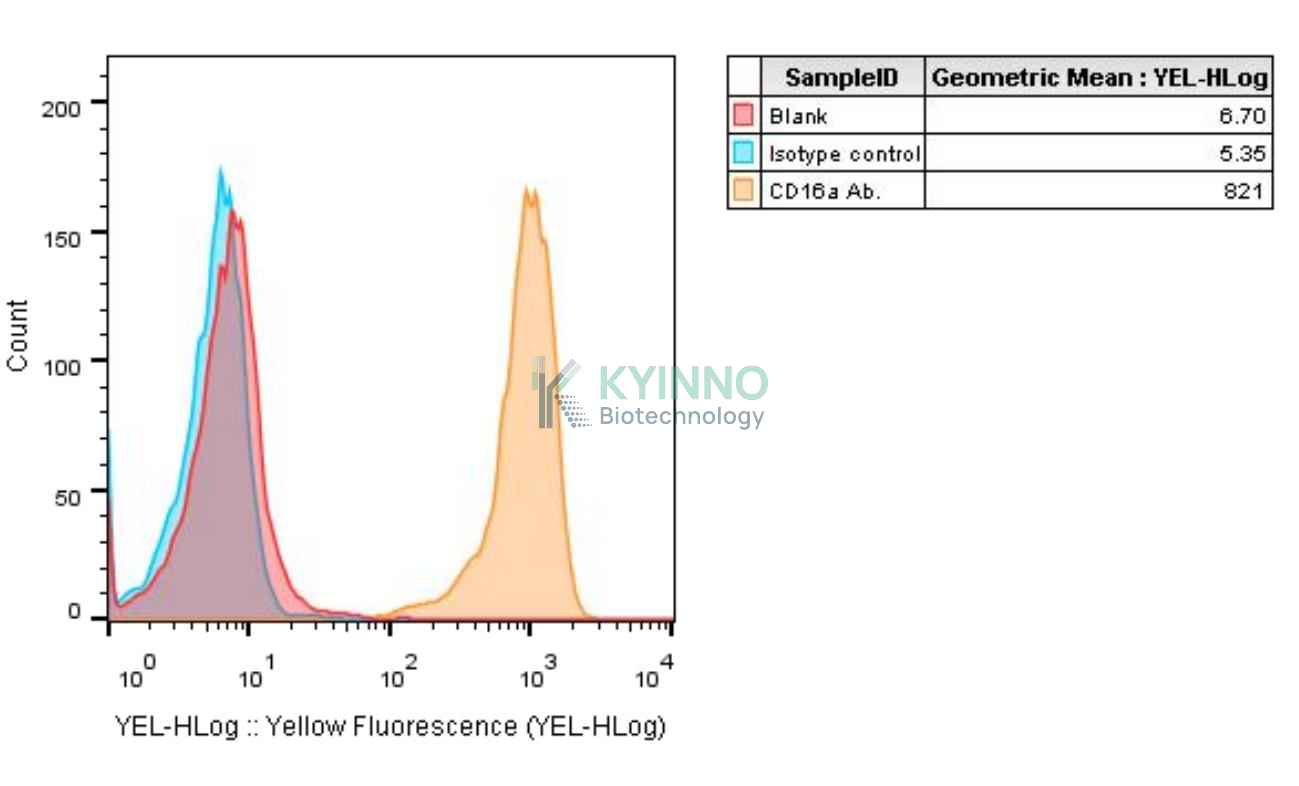
CD16, also named FC gamma RIII, is a low or intermediate affinity FC receptor, and has been identified as two receptors including FcγRIIIa (CD16a) and FcγRIIIb (CD16b). It is involved in phagocytosis, secretion of enzymes and inflammatory mediators, antibody-dependent cytotoxicity and clearance of immune complexes. NFAT proteins, which are expressed in most immune-system cells, play a pivotal role in the transcription of cytokine genes and other genes critical for the immune response. Nuclear factor of activated T cells (NFAT), which is the pharmacological target of immunosuppressants cyclosporine and tacrolimus, has been shown to play an important role not only in T cells (immune system), from which their ame is derived, but also in many biological events. The activity of NFAT proteins is tightly regulated by the calcium/calmodulin-dependent phosphatase calcineurin, a primary target for inhibition by cyclosporin A and FK506. Calcineurin controls the translocation of NFAT proteins from the cytoplasm to the nucleus of activated cells by interacting with an N-terminal regulatory domain conserved in the NFAT family. The DNA-binding domains of NFAT proteins resemble those of Rel-family proteins, and Rel and NFAT proteins show some overlap in their ability to bind to certain regulatory elements in cytokine genes. NFAT is also notable for its ability to bind cooperatively with transcription factors of the AP-1 (Fos/Jun) family to composite NFAT: AP-1 sites, found in the regulatory regions of many genes that are inducibly transcribed by immune-system cells.

Specifications

Catalog NumberKC-1507
Cell Line NameJurkat-NFAT- Luc2-CD16a-V158 Cell Line
Clone Number3#
Host Cell LineJurkat-NFAT-Luc2 cell line
DescriptionJurkat cell line stable expressing exogenous luciferase gene under the control of NFAT responsive element and human FCGR3A amino acid bearing F158V mutation
QuantityTwo vials of frozen cells (≥2-106/vial)
StabilityStable in culture over a minimum of 10 passages
ApplicationDrug screening and biological assays
Freezing Medium70% RPMI1640+20% FBS+10% DMSO
Propagation MediumRPMI1640+10%FBS+300µg/mL Hygromycin B+0.75µg/mL Puromycin
Selection MarkerPuromycin, HygromycinB
MorphologyLymphoblast
SubcultureSplit saturated culture 1:4-1:6 every 2-3 days; seed out at about 1-3 × 10⁵ cells/mL
Incubation37 °C with 5% CO2
StorageLiquid nitrogen immediately upon receiving
Doubling TimeApproximately 26 hours
Mycoplasma StatusNegative
In Vivo ValidationNA

Cell Line Generation

Jurkat NFAT-Luc2-CD16a-F158 reporter cell line was generated using lentiviral vector expressing human CD16a-V158 sequence.

Characterization

Figure 1: Characterization of CD16a-V158 overexpression in Jurkat NFAT-Luc2-CD16a-V158 reporter cell line using FACS.

Figure 2: Characterization of CD16a-V158 overexpression in Jurkat NFAT-Luc2-CD16a-V158 reporter cell line using RT-PCR sequencing.

Figure 3: Jurkat NFAT-Luc2-CD16a-V158 cells and SK-BR-3 cells were seeded into 96-well plates, treated with Herceptin for 6 hours, and then read out using Bright-Glo Detection System.

Figure 4: Jurkat NFAT-Luc2-CD16a-V158 cells and NCI-N87-CLDN18.2 cells were seeded into 96-well plates, treated with MAb362 for 6 hours, and then read out using Bright-Glo Detection System.

Cell Resuscitation

  1. Prewarm culture medium (RPMI1640 + 10% FBS + 300µg/mL Hygromycin B + 0.75µg/mL Puromycin)in a 37°C water bath.
  2. Thaw the frozen vial in a 37°C water bath for 1-2 minutes.
  3. Transfer the vial into biosafety cabinet, and wipe the surface with 70% ethanol.
  4. Unscrew the top of the vial and transfer the cell suspension gently into a sterile centrifuge tube containing 9.0mL complete culture medium.
  5. Spin at ~ 125 × g for 5-7 minutes at room temperature, and discard the supernatant without disturbing the pellet.
  6. Resuspend cell pellet with the appropriate volume of complete medium and transfer the cell suspension into a T25 culture flask.
  7. Incubate the flask at 37°C, 5% CO2 incubator.
  8. Split saturated culture 1:4-1:6 every 2-3 days; seed out at about 1-3 × 10⁵ cells/mL.

Cell Freezing

  1. Prepare the freezing medium (70% RPMI-1640 + 20% FBS + 10% DMSO) fresh immediately before use.
  2. Keep the freezing medium on ice and label cryovials.
  3. Transfer cells to a sterile, conical centrifuge tube, and count the cells.
  4. Centrifuge the cells at 250×g for 5 minutes at room temperature and carefully aspirate off the medium.
  5. Resuspend the cells at a density of at least 3×10⁶ cells/mL in chilled freezing medium.
  6. Aliquot 1 mL of the cell suspension into each cryovial.
  7. Freeze cells in the CoolCell freezing container overnight in a -80°C freezer.
  8. Transfer vials to liquid nitrogen for long-term storage

References

  1. Tada, Minoru, Akiko Ishii-Watabe, Takuo Suzuki, and Nana Kawasaki. 2014. ¡ùDevelopment of a Cell-Based Assay Measuring the Activation of FcÎÛRIIa for the Characterization of Therapeutic Monoclonal Antibodies.¡ì Edited by Paul Zhou. PLoS ONE 9 (4): e95787ÿ89. doi:10.1371/journal.pone.0095787.
  2. Koene, H R, M Kleijer, J Algra, D Roos, A E von dem Borne, and M de Haas. 1997. ¡ùFc gammaRIIIa-158V/F Polymorphism Influences the Binding of IgG by Natural Killer Cell Fc gammaRIIIa, Independently of the Fc gammaRIIIa-48L/R/H Phenotype..¡ì Blood 90 (3): 1109ÿ14.
  3. Rao A, Luo C, Hogan PG. Transcription factors of the NFAT family: regulation and function. Annu Rev Immunol. 1997;15:707-47.
  4. Kitamura N, Kaminuma O. Isoform-Selective NFAT Inhibitor: Potential Usefulness and Development. Int J Mol Sci. 2021 Mar 8;22(5):2725.
  5. Sana I, Mantione ME, Angelillo P, Muzio M. Role of NFAT in Chronic Lymphocytic Leukemia and Other B-Cell Malignancies. Front Oncol. 2021 Apr 1;11:651057.
Please enable JavaScript in your browser to complete this form.