KC-4225

Jurkat-NFAT-Luc2-CD3D-CD3E-KO-2A3-Cell-Line

×
请在浏览器中启用JavaScript来完成此表单。
43406
Home » Jurkat-NFAT-Luc2-CD3D-CD3E-KO-2A3-Cell-Line

Background of Jurkat-NFAT-Luc2-CD3D-CD3E-KO-2A3-Cell-Line

The protein encoded by CD3D is part of the T-cell receptor/CD3 complex (TCR/CD3 complex) and is involved in T-cell development and signal transduction. The encoded membrane protein represents the delta subunit of the CD3 complex, and along with four other CD3 subunits, binds either TCR alpha/beta or TCR gamma/delta to form the TCR/CD3 complex on the surface of T-cells. Defects in this gene are a cause of severe combined immunodeficiency autosomal recessive T-cell-negative/B-cell-positive/NK-cell-positive (SCIDBNK). The protein encoded by CD3E is the CD3-epsilon polypeptide, which together with CD3-gamma, -delta and -zeta, and the T-cell receptor alpha/beta and gamma/delta heterodimers, forms the T-cell receptor-CD3 complex. This complex plays an important role in coupling antigen recognition to several intracellular signal-transduction pathways. The genes encoding the epsilon, gamma and delta polypeptides are located in the same cluster on chromosome 11. The epsilon polypeptide plays an essential role in T-cell development. Defects in this gene cause immunodeficiency.

Specifications

Catalog NumberKC-4225
Cell Line NameJurkat-NFAT-Luc2-CD3D-CD3E-KO-2A3-Cell-Line
Host Cell LineJurkat-NFAT-Luc2-CD3D-KO-1B1
DescriptionStable Jurkat-NFAT-Luc2-CD3D-KO clone with human CD3E gene knockout, No.2A3
QuantityOne vial of frozen cells (≥2-106/vial)
StabilityStable in culture over a minimum of 10 passages
ApplicationDrug screening and biological assays
Freezing MediumRPMI1640+20% FBS+10% DMSO
Propagation MediumRPMI1640+10% FBS+300μg/mL Hygromycin
Selection MarkerN/A
MorphologyLymphoblast
SubcultureSplit saturated culture 1:4-1:6 every 2-3 days; seed out at about 1-3 × 10⁵ cells/mL
Incubation37 °C with 5% CO2
StorageLiquid nitrogen immediately upon receiving
Doubling TimeApproximately 26 hours
Mycoplasma StatusNegative
In Vivo ValidationNA

Cell Line Generation

Jurkat-NFAT-Luc2-CD3D-CD3E-KO-2A3 cell line was generated using the CRISPR method.

Characterization

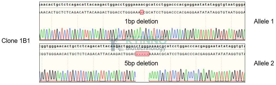
Figure 1: Characterization of CD3D knockout in Jurkat-NFAT-Luc2 using PCR sequencing.

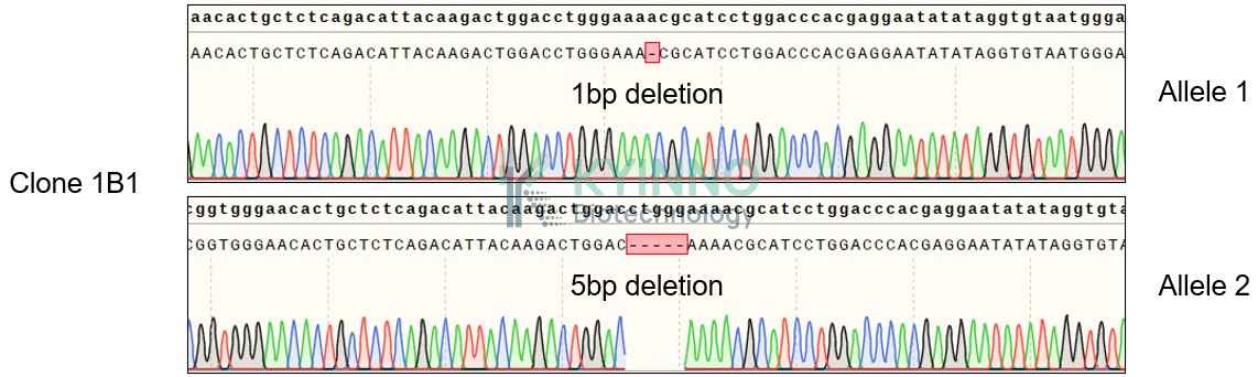
Figure 2: Characterization of CD3D knockout in Jurkat-NFAT-Luc2 using RT-PCR sequencing.

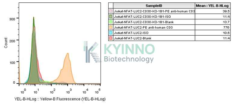
Figure 3: Characterization of CD3D knockout in Jurkat-NFAT-Luc2 using FACS.

Figure 4: Characterization of CD3E knockout in Jurkat-NFAT-Luc2-CD3D-KO using PCR sequencing.

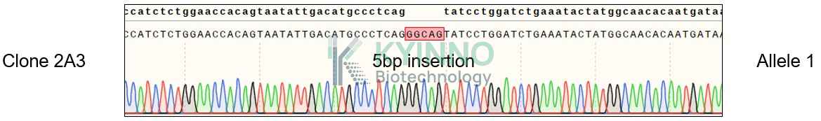
Figure 5: Characterization of CD3E knockout in Jurkat-NFAT-Luc2-CD3D-KO using RT-PCR sequencing.

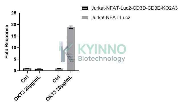
Figure 6: Chareacterization of Jurkat-NFAT-Luc2-CD3-KO clone and Jurkat-NFAT-Luc2 parental cells treated by 20μg/mL OKT3 for 6 hours using Bright-Glo system.

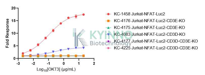
Figure 7. Chareacterization of Jurkat-NFAT-Luc2-CD3-KO clones and Jurkat-NFAT-Luc2 cell line were seeded into the 96-well plate, and treated with OKT3 at a maximum concentration of 20μg/mL for 16 hours, then readout with Bright-Glo system.

Cell Resuscitation

  1. Prewarm culture medium (RPMI1640 + 10% FBS+300μg/mL Hygromycin)in a 37°C water bath.
  2. Thaw the frozen vial in a 37°C water bath for 1-2 minutes.
  3. Transfer the vial into biosafety cabinet, and wipe the surface with 70% ethanol.
  4. Unscrew the top of the vial and transfer the cell suspension gently into a sterile centrifuge tube containing 9.0mL complete culture medium.
  5. Spin at ~ 125 × g for 5-7 minutes at room temperature, and discard the supernatant without disturbing the pellet.
  6. Resuspend cell pellet with the appropriate volume of complete medium and transfer the cell suspension into a T25 culture flask.
  7. Incubate the flask at 37°C, 5% CO2 incubator.
  8. Split saturated culture 1:4-1:6 every 2-3 days; seed out at about 1-3 × 10⁵ cells/mL.

Cell Freezing

  1. Prepare the freezing medium (70% RPMI-1640 + 20% FBS + 10% DMSO) fresh immediately before use.
  2. Keep the freezing medium on ice and label cryovials.
  3. Transfer cells to a sterile, conical centrifuge tube, and count the cells.
  4. Centrifuge the cells at 250×g for 5 minutes at room temperature and carefully aspirate off the medium.
  5. Resuspend the cells at a density of at least 3×10⁶ cells/mL in chilled freezing medium.
  6. Aliquot 1 mL of the cell suspension into each cryovial.
  7. Freeze cells in the CoolCell freezing container overnight in a -80°C freezer.
  8. Transfer vials to liquid nitrogen for long-term storage

References

  1. https://www.ncbi.nlm.nih.gov/gene/915 2.https://www.ncbi.nlm.nih.gov/gene/916

Use License Agreement

Research Use Only.
Not for use in diagnostic procedures or therapeutic applications.
Redistribution of the cell line or its derivatives is prohibited without prior written permission from Kyinno Biotechnology.
请在浏览器中启用JavaScript来完成此表单。