KC-2135

Jurkat-NFAT-Luc2-CTLA4-Cell-Line

×
Please enable JavaScript in your browser to complete this form.
43856
Home » Jurkat-NFAT-Luc2-CTLA4-Cell-Line

Background of Jurkat-NFAT-Luc2-CTLA4-Cell-Line

CTLA4 (CD152) has been in the centre of attention for its pivotal role in autoimmunity and cancer. Although not fully understood, CTLA4 with no doubt plays an important role in the maintenance of the immune response by its expression on activated and regulatory T cells. CTLA4 has many variants and polymorphic forms, some present in regulatory positions, some in 3' UTR and the most important one in the leader sequence (+49 A/G).CTL-associated antigen 4 (CTLA4) is a well-established immune checkpoint for antitumor immune responses. The protumorigenic function of CTLA4 is believed to be limited to T-cell inhibition by countering the activity of the T-cell costimulating receptor CD28.

Specifications

Catalog NumberKC-2135
Cell Line NameJurkat-NFAT-Luc2-CTLA4-Cell-Line
Host Cell LineJurkat
DescriptionStable Jurkat cell line expressing exogenous luciferase gene under the control of CTLA4 signaling pathway
QuantityTwo vials of frozen cells (≥2-106/vial)
StabilityStable in culture over a minimum of 10 passages
ApplicationCell model for monitoring EGFR signaling pathway
Freezing Medium70% RPMI1640+20% FBS+10% DMSO
Propagation MediumRPMI1640+10%FBS+300µg/mL Hygromycin B+0.75µg/mL Puromycin
Selection MarkerPuromycin, HygromycinB
MorphologyLymphoblast
SubcultureSplit saturated culture 1:4-1:5 every 2-3 days; seed out at about 1-3 × 10⁵ cells/mL
Incubation37 °C with 5% CO2
StorageLiquid nitrogen immediately upon receiving
Doubling TimeApproximately 26 hours
Mycoplasma StatusNegative
In Vivo ValidationNA

Cell Line Generation

Jurkat-NFAT-Luc2-CTLA4 cell line was generated using lentivirus expressing EGFR sequence.

Characterization

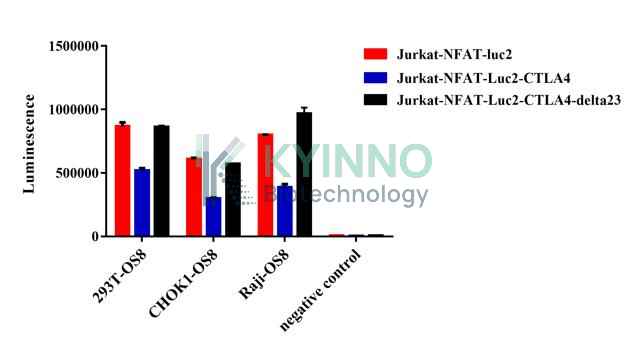
Figure 1. Jurkat-NFAT-Luc2-CTLA4 cell line was seeded into the 96-well plate, and co-culture with 293T-OS8/CHOK1-OS8/Raji-OS8 for 6 hours, then readout with Bright-Glo system.

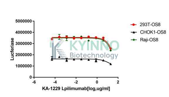
Figure 2. Jurkat-NFAT-Luc2-CTLA4 cell line was seeded into the 96-well plate, and treated with CTLA4 antibody at a maximum concentration 20 μg/mL for 1 hours, and co-culture with 293T-OS8/CHOK1-OS8/Raji-OS8 for 6 hours, then readout with Bright-Glo system.

Cell Resuscitation

  1. Prewarm culture medium (RPMI1640 + 10% FBS + 300µg/mL Hygromycin B + 0.75µg/mL Puromycin)in a 37°C water bath.
  2. Thaw the frozen vial in a 37°C water bath for 1-2 minutes.
  3. Transfer the vial into biosafety cabinet, and wipe the surface with 70% ethanol.
  4. Unscrew the top of the vial and transfer the cell suspension gently into a sterile centrifuge tube containing 9.0mL complete culture medium.
  5. Spin at ~ 125 × g for 5-7 minutes at room temperature, and discard the supernatant without disturbing the pellet.
  6. Resuspend cell pellet with the appropriate volume of complete medium and transfer the cell suspension into a T25 culture flask.
  7. Incubate the flask at 37°C, 5% CO2 incubator.
  8. Split saturated culture 1:4-1:5 every 2-3 days; seed out at about 1-3 × 10⁵ cells/mL.

Cell Freezing

  1. Prepare the freezing medium (70% RPMI1640 + 20% FBS + 10% DMSO) fresh immediately before use.
  2. Keep the freezing medium on ice and label cryovials.
  3. Transfer cells to a sterile, conical centrifuge tube, and count the cells.
  4. Centrifuge the cells at 250×g for 5 minutes at room temperature and carefully aspirate off the medium.
  5. Resuspend the cells at a density of at least 3×10⁶ cells/mL in chilled freezing medium.
  6. Aliquot 1 mL of the cell suspension into each cryovial.
  7. Freeze cells in the CoolCell freezing container overnight in a -80°C freezer.
  8. Transfer vials to liquid nitrogen for long-term storage

References

  1. Ghaderi A. CTLA4 gene variants in autoimmunity and cancer: a comparative review. Iran J Immunol. 2011 Sep;8(3):127-49.
  2. Herrmann A, Lahtz C, Nagao T, Song JY, Chan WC, Lee H, Yue C, Look T, Mülfarth R, Li W, Jenkins K, Williams J, Budde LE, Forman S, Kwak L, Blankenstein T, Yu H. CTLA4 Promotes Tyk2-STAT3-Dependent B-cell Oncogenicity. Cancer Res. 2017 Sep 15;77(18):5118-5128.
Please enable JavaScript in your browser to complete this form.