KC-1503

Jurkat-NFAT-Luc2-PD1 Cell Line

×
Please enable JavaScript in your browser to complete this form.
22267
Home » Jurkat-NFAT-Luc2-PD1 Cell Line

Background of Jurkat-NFAT-Luc2-PD1 Cell Line

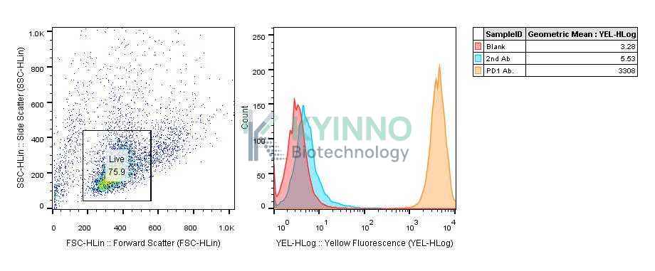
PD-1, human programmed cell death 1 protein, is a receptor belonging to the immunoglobulin superfamily which is expressed primarily on activated T cells. NK cells, B cells, and monocytes. PD-1 interaction with its ligands, PD-L1 and PD-L2, leads to the downregulation of T cell responses, including T cell proliferation and cytokine production, and limits immune destruction of tissues. The interaction between PD-1 and its ligands, thus plays a role in maintaining the balance between immune activation and tolerance, potentially including tumor tolerance. 
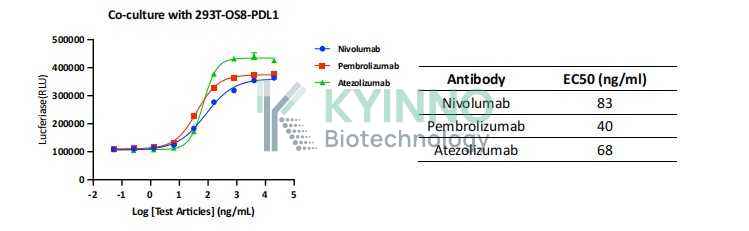
NFAT (nuclear factor of activator T cells) proteins belong to a family of transcription factors that are very important in T cells activation, differentiation, and tolerance. NFAT pathway is activated by Ca2+ and the Ca2+/calmodulin- dependent serine phosphatase calcineurin after the engagement of the T cell receptor (TCR).

Specifications

Catalog NumberKC-1503
Cell Line NameJurkat-NFAT-Luc2-PD1 Cell Line
Host Cell LineJurkat-NFAT-Luc2
DescriptionJurkat cell line stable expressing exogenous luciferase gene under the control of NFAT responsive element and full-length human PD1 protein.
QuantityTwo vials of frozen cells (≥2-106/vial)
StabilityStable in culture over a minimum of 10 passages
ApplicationDrug screening and biological assays
Freezing Medium70% RPMI1640+20% FBS+10% DMSO
Propagation MediumRPMI1640+10% FBS+300ug/ml Hygromycin+0.75ug/ml puromycin
Selection MarkerPuromycin, Hygromycin B
MorphologyLymphoblast
SubcultureSplit saturated culture 1:4-1:5 every 2-3 days; seed out at about 1-3 × 10⁵ cells/mL
Incubation37 °C with 5% CO2
StorageLiquid nitrogen immediately upon receiving
Doubling TimeApproximately 30 hours
Mycoplasma StatusNegative
In Vivo ValidationNA

Cell Line Generation

Jurkat-NFAT-Luc2-PD1 cell line was generated using lentiviral vector expressing full-length sequence.

Characterization

Figure 1. Characterization of PD1 overexpression in Jurkat-NFAT-Luc2-PD1 reporter cell line.

Figure 2. Jurkat-NFAT-Luc2-PD1 cells and KC-1148 293T-OS8-PDL1 cells were seeded into 96-well plates, treated with different antibodies for 6 hours, and then read out using Bright-Glo Detection System.

Cell Resuscitation

  1. Prewarm culture medium (RPMI1640 supplemented with 10% FBS, 300μg/ml Hygromycin and 0.75μg/ml Puromycin)in a 37°C water bath.
  2. Thaw the frozen vial in a 37°C water bath for 1-2 minutes.
  3. Transfer the vial into biosafety cabinet, and wipe the surface with 70% ethanol.
  4. Unscrew the top of the vial and transfer the cell suspension gently into a sterile centrifuge tube containing 9.0mL complete culture medium.
  5. Spin at ~ 125 × g for 5-7 minutes at room temperature, and discard the supernatant without disturbing the pellet.
  6. Resuspend cell pellet with the appropriate volume of complete medium and transfer the cell suspension into a T25 culture flask.
  7. Incubate the flask at 37°C, 5% CO2 incubator.
  8. Split saturated culture 1:4-1:5 every 2-3 days; seed out at about 1-3 × 10⁵ cells/mL.

Cell Freezing

  1. Prepare the freezing medium (70% 1640 + 20% FBS + 10% DMSO) fresh immediately before use.
  2. Keep the freezing medium on ice and label cryovials.
  3. Transfer cells to a sterile, conical centrifuge tube, and count the cells.
  4. Centrifuge the cells at 250×g for 5 minutes at room temperature and carefully aspirate off the medium.
  5. Resuspend the cells at a density of at least 3×10⁶ cells/mL in chilled freezing medium.
  6. Aliquot 1 mL of the cell suspension into each cryovial.
  7. Freeze cells in the CoolCell freezing container overnight in a -80°C freezer.
  8. Transfer vials to liquid nitrogen for long-term storage

References

  1. Boussiotis, V. A. Molecular and Biochemical Aspects of the PD-1 Checkpoint Pathway. N Engl J Med 375, 1767– 1778 (2016).
Please enable JavaScript in your browser to complete this form.